. 2016:2016:7521478.
doi: 10.1155/2016/7521478. Epub 2016 Mar 23.

The Diagnostic Accuracy of Optical Coherence Tomography Angiography for Neovascular Age-Related Macular Degeneration: A Comparison with Fundus Fluorescein Angiography

Affiliations

The Diagnostic Accuracy of Optical Coherence Tomography Angiography for Neovascular Age-Related Macular Degeneration: A Comparison with Fundus Fluorescein Angiography

Jingwen Gong et al. J Ophthalmol. 2016.

Abstract

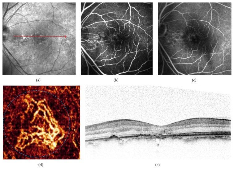
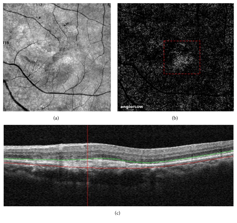
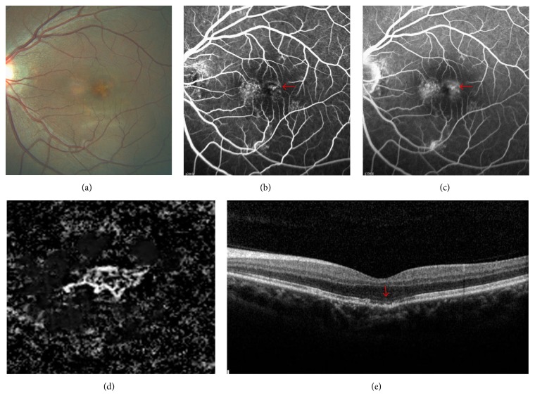
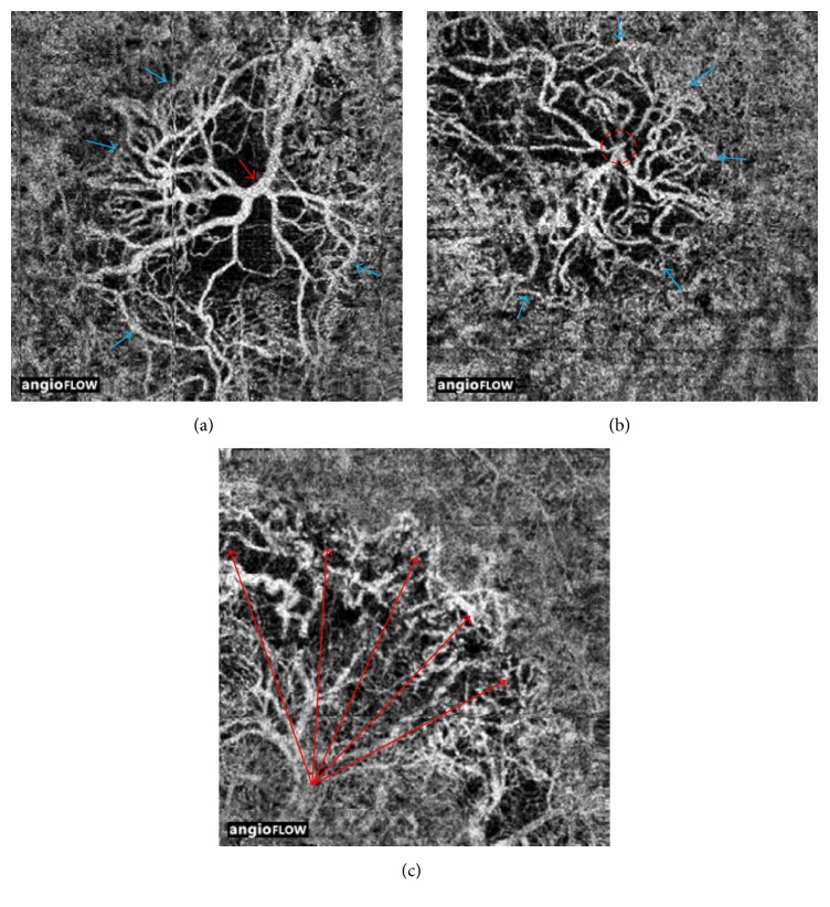
Purpose. To describe the morphological characteristics and efficacy of OCTA in detecting CNV in nAMD. We retrospectively reviewed 53 patients (86 eyes) with suspected CNV secondary to wet AMD. All the patients underwent a multimodal assessment for CNV. Two independent readers calculated the sensitivity and specificity of OCTA in detecting CNV compared with FA. A qualitative analysis of OCTA was also performed to describe the morphological appearance of CNV. Among 86 eyes of 53 patients, 52 eyes were diagnosed as having CNV based on the FA imaging analysis. According to FA, CNV was classified as classic in 28 eyes, predominantly classic in 6 eyes, minimally classic in 9 eyes, and occult in 9 eyes. In 56 eyes, CNV was visualized on OCTA and corresponding OCT B-scans. In total, 46.4% (26/56) had well-circumscribed vessels, and 53.6% (30/56) showed poorly circumscribed vessels. There were 11 false positives and 7 false negatives using OCTA. The specificity of OCTA for the detection of CNV was 67.6%, with sensitivity of 86.5%. OCTA may help in the noninvasive diagnosis of CNV and may provide a method for monitoring the evolution of CNV.


Figures

Figure 1
Figure 1
Images of choroidal neovascularization (CNV) observed in false-positive case on optical coherence tomography angiography (OCTA). (a) IR imaging from a 55-year-old man who was previously treated with 3 intravitreal anti-VEGF injections. (b and c) Early- and late-frame FA images of the patient displaying mild hyperfluorescence that is stable throughout the FA in the region of CNV without pooling. (d) A 33-mm En face angiogram of the outer retina showing a well-circumscribed CNV in the subfoveal (blue dashed circle). (e) Intraretinal cystic spaces and mild RPE irregularity seen on the spectral-domain optical coherence tomography occur at matching locations on IR imaging (red line).
Figure 2
Figure 2
Distinguishing the choriocapillaris artifact from choroidal neovascularization (CNV). (a and b) A 66-mm En face angiogram of the outer retina from a 62-year-old woman who were previously treated with 18 intravitreal anti-VEGF injections showing a homogenous and ill-defined network of vessels with a granular appearance (red dashed box). (c) Spectral-domain optical coherence tomography displaying a fusiform or spindle-shaped complex of high reflectivity located within the subretinal space.
Figure 3
Figure 3
Images of subretinal hemorrhage observed in false-negative case on optical coherence tomography angiography (OCTA). (a) Color fundus photograph of the patient showing large amounts of subretinal hemorrhage. (b) Late-frame FA image displaying big leakage and pooling at the inferonasal to the macula together with posterior pole pigment epithelium detachment, also with masking effect attributable to subretinal hemorrhage. (c) Spectral-domain optical coherence tomography (SD-OCT) demonstrating large amounts of subretinal hemorrhage over the retinal pigment epithelial (RPE) detachment.
Figure 4
Figure 4
Multimodal imaging of neovascularization in age-related macular degeneration. (a) A color fundus photograph image from a 50-year-old male patient who complained of the reduced vision for one week displaying the presence of drusen in the subfoveal. (b) Early-frame FA showing hyperfluorescence in the region of CNV (red arrow). (c) Late-frame FA image showing leakage and pooling (red arrow). (d) A 33-mm En face angiogram of the outer retina demonstrating a small well-circumscribed CNV in the subfoveal surrounded by a halo of choriocapillaris vessels. (e) Spectral-domain optical coherence tomography (SD-OCT) only showing drusen, disrupted of retinal pigment epithelium (RPE), and small defect of the photoreceptor layer which were graded as dry AMD (red arrow).
Figure 5
Figure 5
Optical coherence tomography angiography (OCTA) of treated choroidal neovascularization. (a) A 33-mm OCTA en face projection image from an 84-year-old man who was treated with 20 intravitreal anti-VEGF injections. Note the large diameter of the trunk vessels (red arrow). At the periphery there appears to be a defined anastomotic connection around the border (blue arrows). (b) A 33-mm En face angiogram of the neovascular membrane from a 65-year-old man who were previously treated with 18 intravitreal anti-VEGF injections showing the red dashed circle that encompasses the trunk vessels of the lesion. Note the neovascular complex with vessels radiating in all directions from the center and the terminal loops of the vessels (blue arrows). (c) A 33-mm OCTA en face projection image from a 65-year-old man who were treated with 16 intravitreal anti-VEGF injections. Note the long extent of the vessels as they reach the edge of the lesion (red arrows).

References

    1. Ferris F. L., III, Fine S. L., Hyman L. Age-related macular degeneration and blindness due to neovascular maculopathy. Archives of Ophthalmology. 1984;102(11):1640–1642. doi: 10.1001/archopht.1984.01040031330019. - DOI - PubMed
    1. Ambati J., Ambati B. K., Yoo S. H., Ianchulev S., Adamis A. P. Age-related macular degeneration: etiology, pathogenesis, and therapeutic strategies. Survey of Ophthalmology. 2003;48(3):257–293. doi: 10.1016/s0039-6257(03)00030-4. - DOI - PubMed
    1. Do D. V. Detection of new-onset choroidal neovascularization. Current Opinion in Ophthalmology. 2013;24(3):224–227. doi: 10.1097/ICU.0b013e32835fd7dd. - DOI - PubMed
    1. Stanga P. E., Lim J. I., Hamilton P. Indocyanine green angiography in chorioretinal diseases: indications and interpretation: an evidence-based update. Ophthalmology. 2003;110(1):15–50. doi: 10.1016/s0161-6420(02)01563-4. - DOI - PubMed
    1. López-Sáez M. P., Ordoqui E., Tornero P., et al. Fluorescein-induced allergic reaction. Annals of Allergy, Asthma and Immunology. 1998;81(5 I):428–430. doi: 10.1016/S1081-1206(10)63140-7. - DOI - PubMed

LinkOut - more resources