

Neonatal Abdominal Hemangiomatosis: Propranolol beyond Infantile Hemangioma
- PMID: 27110421
- PMCID: PMC4826694
- DOI: 10.1155/2016/9803975
Neonatal Abdominal Hemangiomatosis: Propranolol beyond Infantile Hemangioma
Abstract
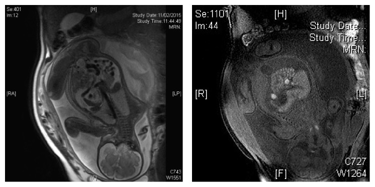
Hemangioma is the most common vascular tumor of infancy; presentation is often as cutaneous infantile hemangioma (IH). Cutaneous hemangioma is a clinical diagnosis. Most IHs follow a benign course, with complete involution without treatment in the majority of cases. Visceral hemangioma often involves the liver and manifests as a life-threatening disorder. Hepatic hemangiomas may be associated with high output cardiac failure, coagulopathy, and hepatomegaly which generally develop between 1 and 16 weeks of age. Mortality has been reportedly high without treatment. We report a rare case of a male infant with neonatal hemangiomatosis with diffuse peritoneal involvement, which mimicked a malignant-looking tumor on imaging, and discuss therapeutic options and efficacy. Propranolol is efficacious for IH but generally not useful for other forms of vascular hemangiomas, tumors, and malformations. In our case of neonatal peritoneal hemangiomatosis, propranolol appears to have halted the growth and possibly expedite the involution of the hemangiomatosis without other treatments.
Figures


Similar articles
-
Diffuse neonatal hemangiomatosis in a very low-birthweight infant treated with erythropoietin.Pediatr Int. 2015 Apr;57(2):e34-6. doi: 10.1111/ped.12517. Pediatr Int. 2015. PMID: 25868957
-
Efficacy of intravenous propranolol for life-threatening diffuse neonatal hemangiomatosis.Pediatr Dermatol. 2022 Jul;39(4):613-615. doi: 10.1111/pde.15028. Epub 2022 May 16. Pediatr Dermatol. 2022. PMID: 35575222
-
Diffuse neonatal hemangiomatosis: an evidence-based review of case reports in the literature.J Am Acad Dermatol. 2012 Nov;67(5):898-903. doi: 10.1016/j.jaad.2012.01.018. Epub 2012 Feb 15. J Am Acad Dermatol. 2012. PMID: 22341467
-
Infantile hemangiomas, complications and treatments.Semin Cutan Med Surg. 2016 Sep;35(3):108-16. doi: 10.12788/j.sder.2016.050. Semin Cutan Med Surg. 2016. PMID: 27607318 Review.
-
Multiple cutaneous and hepatic infantile hemangiomas having a successful response to propranolol as monotherapy at neonatal period.G Ital Dermatol Venereol. 2013 Oct;148(5):525-30. G Ital Dermatol Venereol. 2013. PMID: 24005146 Review.
Cited by
-
Diffuse neonatal hemangiomatosis presenting as congestive heart failure.Dermatol Pract Concept. 2017 Jul 31;7(3):66-69. doi: 10.5826/dpc.0703a15. eCollection 2017 Jul. Dermatol Pract Concept. 2017. PMID: 29085724 Free PMC article.
-
From Severe Anemia to Intestinal Hemangiomatosis, a Bumpy Road-A Case Report and Literature Review.Diagnostics (Basel). 2024 Jan 31;14(3):310. doi: 10.3390/diagnostics14030310. Diagnostics (Basel). 2024. PMID: 38337828 Free PMC article.
-
Recurrent bloody stools associated with visceral infantile haemangioma in a preterm twin girl.BMJ Case Rep. 2018 Dec 3;11(1):bcr2018226564. doi: 10.1136/bcr-2018-226564. BMJ Case Rep. 2018. PMID: 30567166 Free PMC article.
-
Infantile Hemangiomas: An Update on Pathogenesis and Treatment.J Clin Med. 2021 Oct 9;10(20):4631. doi: 10.3390/jcm10204631. J Clin Med. 2021. PMID: 34682753 Free PMC article. Review.
-
Vascular anomalies associated with hepatic shunting.World J Gastroenterol. 2020 Nov 14;26(42):6582-6598. doi: 10.3748/wjg.v26.i42.6582. World J Gastroenterol. 2020. PMID: 33268948 Free PMC article. Review.
References
-
- Zhao F.-Y., Gao Y., Wu M.-J., Luo Q.-F., Liu Y., Xu Z.-Q. Diagnosis and therapy on hemangiomas and vascular malformation in view of the new classification. Beijing Da Xue Xue Bao. 2009;41(1):21–27. - PubMed
-
- Hon K. L. E., Shen P. C. H., Li J. J. X., Chow C. M., Luk D. C. K. Pediatric vascular anomalies: an overview of management. Clinical Medicine Insights: Dermatology. 2014;7:1–7.
LinkOut - more resources
Full Text Sources
Other Literature Sources
