Review
. 2016 Apr 21;8(4):112.
doi: 10.3390/v8040112.

Measles Virus Fusion Protein: Structure, Function and Inhibition

Affiliations
Review

Measles Virus Fusion Protein: Structure, Function and Inhibition

Philippe Plattet et al. Viruses. .

Abstract

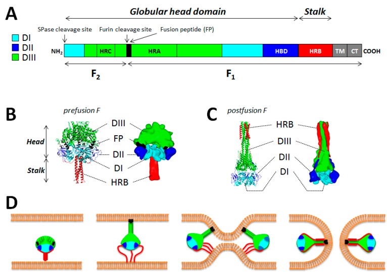
Measles virus (MeV), a highly contagious member of the Paramyxoviridae family, causes measles in humans. The Paramyxoviridae family of negative single-stranded enveloped viruses includes several important human and animal pathogens, with MeV causing approximately 120,000 deaths annually. MeV and canine distemper virus (CDV)-mediated diseases can be prevented by vaccination. However, sub-optimal vaccine delivery continues to foster MeV outbreaks. Post-exposure prophylaxis with antivirals has been proposed as a novel strategy to complement vaccination programs by filling herd immunity gaps. Recent research has shown that membrane fusion induced by the morbillivirus glycoproteins is the first critical step for viral entry and infection, and determines cell pathology and disease outcome. Our molecular understanding of morbillivirus-associated membrane fusion has greatly progressed towards the feasibility to control this process by treating the fusion glycoprotein with inhibitory molecules. Current approaches to develop anti-membrane fusion drugs and our knowledge on drug resistance mechanisms strongly suggest that combined therapies will be a prerequisite. Thus, discovery of additional anti-fusion and/or anti-attachment protein small-molecule compounds may eventually translate into realistic therapeutic options.

Keywords: cell entry; fusion protein; inhibitors and mechanisms of adaptation; measles virus; membrane fusion; neuroinvasion; structural changes.


Figures

Figure 1
Figure 1
Structure/function of the measles virus (MeV) fusion protein. (A) The important functional domains of F are color-coded. The F1 and F2 subunits are shown. While the DI (light blue), DII (dark blue) and DIII (green) domains define the globular head domain, the HRB region defines the stalk. TM: transmembrane domain; CT: cytoplasmic tail; HRA-C: heptad repeat region A–C; FP: fusion peptide; HBD: H-binding domain; SPase: signal peptidase. (B and C) Structural differences between the pre and postfusion states of F. Images were obtained from homology models performed with the canine distemper virus (CDV) F sequence (strain A75/17) and based on the atomic coordinates of the human parainfluenza virus type 5 (hPIV5) F prefusion state (PDB 2B9B) and hPIV3 F postfusion state (PDB 1ZTM). DI, DII and DIII are color-coded as indicated in (A). Images were generated using either the PyMol (high resolution) or Sculptor (low resolution) software packages (D) Model of membrane fusion induced by measles virus F. Once activated by H, prefusion F may disengage from H interaction. Then the HRB region of the stalk may melt, which consequently may enable the 11 segments positioned in the globular head to rearrange into a long three-helix bundle (3HB) coiled-coil. This would in turn propel the fusion peptide at the top of the 3HB and allow its insertion into the target plasma membrane. This prehairpin intermediate then collapses, which brings both membranes into close proximity. Finally, F may reach the highly stable postfusion state, which carries the six-helix bundle (6HB) formed by HRA and HRB segments. This stage is thought to correlate with membrane merging and ensuing fusion pore opening. This model likely requires the concerted action of more than one F molecules. This model has been described first by Yin and colleagues [79,86].
Figure 2
Figure 2
Possible structure(s) of the MeV attachment protein. (A and B) Crystal structures of MeV H in complex with the receptor signaling lymphocyte activation molecule (SLAM) (not shown). Two tetrameric configurations were determined (PDB 3ALZ and 3ALX). High-resolution structures were morphed into low-resolution images using the Sculptor software package; (C and D) Two alternative folds of H tetramers. Images were obtained from homology models performed with the canine distemper virus CDV) H sequence (strain A75/17) and based on the atomic coordinates of Newcastle disease virus (NDV) HN (PDB 3T1E) and hPIV5 HN (PDB 4JF7). The dimers of the attachment protein tetramer are color-coded in green and red.
Figure 3
Figure 3
Model of MeV H/F assembly. (A) Representation of MeV F as a trimer (upper panel) or monomer (lower panel) with the DI, DII and DIII domains color-coded as indicated in Figure 1A. The highlighted box shows a zoom of the Ig-like domain proposed to be important for interaction with H (HBD). The two key hydrophobic residues proposed (in CDV F) to control H interaction are represented as spheres and color-coded in red (l506 and K508) [133]. Images were generated using the PyMol software package (V 1.0, DeLano Scientific, Palo Alto, California, USA); (B) Putative H/F assembly. The presumptive HBD of one F-monomer is aligned with the proposed H-stalk segment involved in a short range interaction with F (FBD). The “spacer” H-stalk module, which enables positioning of the two dimeric head units above the F trimer, is represented as a box.
Figure 4
Figure 4
Models of F activation by paramyxovirus attachment proteins. (AE) Latest models have proposed how attachment protein of different members of the Paramyxovirinae subfamily activates F as a consequence of attachment protein-receptor binding. Importantly, all models predict a receptor-induced overall re-positioning of the head regions upon receptor engagements, which may then act as a universal core mechanism for F activation (first described by Bose and colleagues [122]). H tetramers are colored in green. Pre-activated F and activated F are highlighted in blue and red, respectively. In model (D) (bi-dentate model) the conformational changes occurring within the head domain prior to heads’ re-positioning are represented by oval structures. The attachment protein stalk regions that are implicated in F activation are shown as black lines (models BD).

Similar articles

Cited by

References

    1. Lamb R.A., Parks G.D. Paramyxoviridae: The viruses and their replication. In: Fileds B., Knipe D.M., Howley P.M., editors. Fields’ Virology. 5th ed. Lippincott Williams & Wilkins; Philadelphia, PA, USA: 2007. pp. 1449–1496.
    1. Simons E., Ferrari M., Fricks J., Wannemuehler K., Anand A., Burton A., Strebel P. Assessment of the 2010 global measles mortality reduction goal: Results from a model of surveillance data. Lancet. 2012;379:2173–2178. doi: 10.1016/S0140-6736(12)60522-4. - DOI - PubMed
    1. Plemper R.K., Hammond A.L. Synergizing vaccinations with therapeutics for measles eradication. Expert Opin. Drug Discov. 2014;9:201–214. doi: 10.1517/17460441.2014.867324. - DOI - PMC - PubMed
    1. Chua K.B., Bellini W.J., Rota P.A., Harcourt B.H., Tamin A., Lam S.K., Ksiazek T.G., Rollin P.E., Zaki S.R., Shieh W., et al. Nipah virus: A recently emergent deadly paramyxovirus. Science. 2000;288:1432–1435. doi: 10.1126/science.288.5470.1432. - DOI - PubMed
    1. Westbury H.A., Hooper P.T., Selleck P.W., Murray P.K. Equine morbillivirus pneumonia: Susceptibility of laboratory animals to the virus. Aust. Vet. J. 1995;72:278–279. doi: 10.1111/j.1751-0813.1995.tb03549.x. - DOI - PubMed

Publication types

MeSH terms