. 2016 Jul;125(1):147-58.
doi: 10.1097/ALN.0000000000001134.

Effects of γ-Aminobutyric Acid Type A Receptor Modulation by Flumazenil on Emergence from General Anesthesia

Affiliations

Effects of γ-Aminobutyric Acid Type A Receptor Modulation by Flumazenil on Emergence from General Anesthesia

Seyed A Safavynia et al. Anesthesiology. 2016 Jul.

Abstract

Background: Transitions into conscious states are partially mediated by inactivation of sleep networks and activation of arousal networks. Pharmacologic hastening of emergence from general anesthesia has largely focused on activating subcortical monoaminergic networks, with little attention on antagonizing the γ-aminobutyric acid type A receptor (GABAAR). As the GABAAR mediates the clinical effects of many common general anesthetics, the authors hypothesized that negative GABAAR modulators would hasten emergence, possibly via cortical networks involved in sleep.

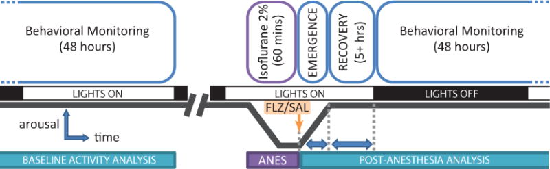
Methods: The authors investigated the capacity of the benzodiazepine rescue agent, flumazenil, which had been recently shown to promote wakefulness in hypersomnia patients, to alter emergence. Using an in vivo rodent model and an in vitro GABAAR heterologous expression system, they measured flumazenil's effects on behavioral, neurophysiologic, and electrophysiologic correlates of emergence from isoflurane anesthesia.

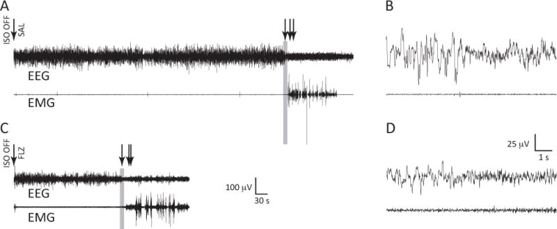
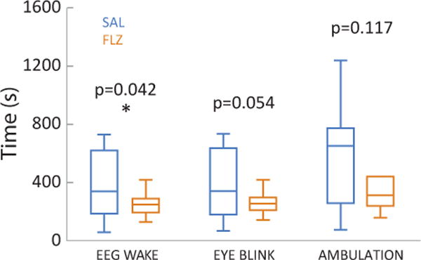
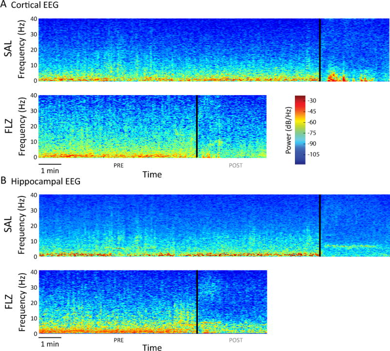
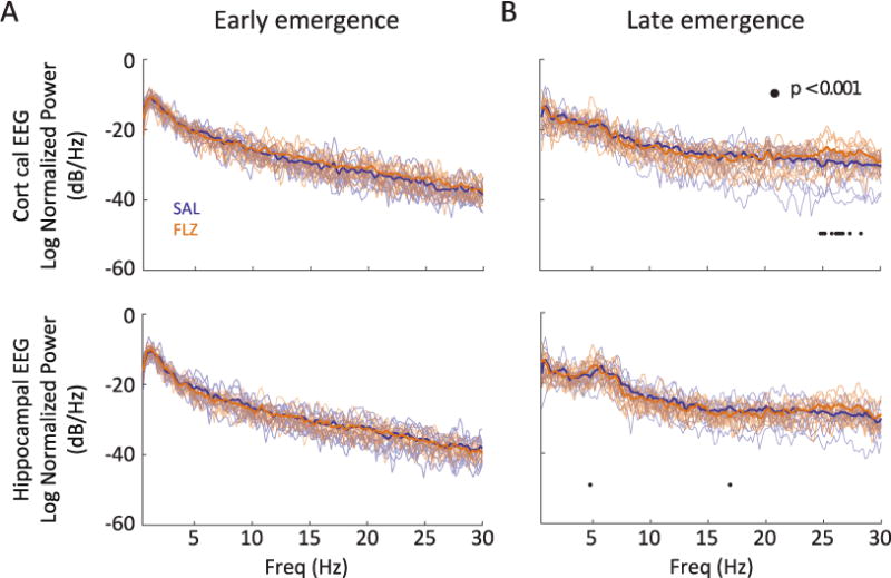
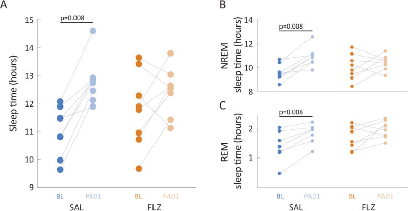
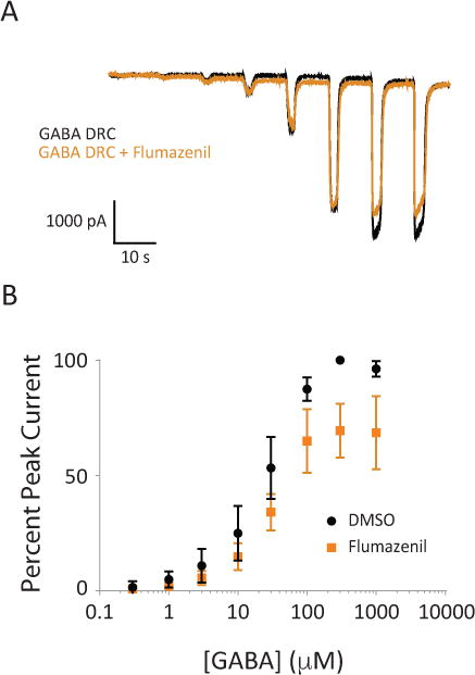
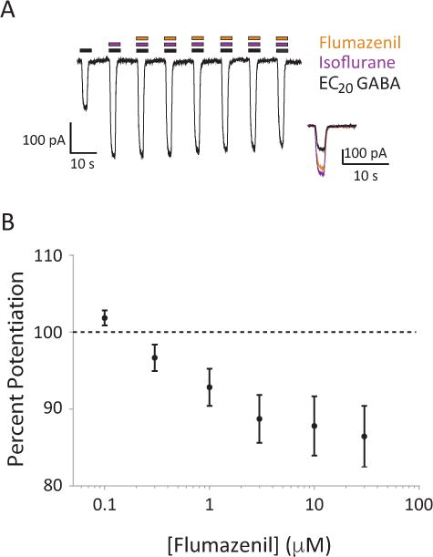
Results: Animals administered intravenous flumazenil (0.4 mg/kg, n = 8) exhibited hastened emergence compared to saline-treated animals (n = 8) at cessation of isoflurane anesthesia. Wake-like electroencephalographic patterns occurred sooner and exhibited more high-frequency electroencephalography power after flumazenil administration (median latency ± median absolute deviation: 290 ± 34 s) compared to saline administration (473 ± 186 s; P = 0.042). Moreover, in flumazenil-treated animals, there was a decreased impact on postanesthesia sleep. In vitro experiments in human embryonic kidney-293T cells demonstrated that flumazenil inhibited isoflurane-mediated GABA current enhancement (n = 34 cells, 88.7 ± 2.42% potentiation at 3 μM). Moreover, flumazenil exhibited weak agonist activity on the GABAAR (n = 10 cells, 10.3 ± 3.96% peak GABA EC20 current at 1 μM).

Conclusions: Flumazenil can modulate emergence from isoflurane anesthesia. The authors highlight the complex role GABAARs play in mediating consciousness and provide mechanistic links between emergence from anesthesia and arousal.


Figures

Figure 1
Figure 1
Experimental paradigm. Adult male rats were outfitted with EEG/EMG electrodes and EEG/EMG was recorded for a 48-hour period prior to anesthesia induction for sleep scoring. Animals were then administered an induction dose of isoflurane, followed by a maintenance dose of 2% isoflurane in oxygen for 60 minutes. At the cessation of isoflurane, intravenous flumazenil (0.4 mg/kg) or an equivalent volume of saline was administered, and the animal was placed into the long-term EEG/EMG recording chamber. Emergence characteristics (EEG wake, eye blink, ambulation) were measured. EEG/EMG were recorded for at least 48 hours postoperatively for qEEG analysis and sleep scoring.
Figure 2
Figure 2
EEG/EMG recordings of the transition to arousal from anesthesia. A, Representative waveforms from an animal given saline (SAL) via tail vein catheter at the cessation of isoflurane anesthesia. EEG from the medial (hippocampal) lead is shown. Arrows designate (from left to right): isoflurane cessation and administration of saline, EEG wake (defined as a transition to lower amplitude, faster oscillatory activity), eye blink, and ambulation. B, Time-expanded 10-second epoch capturing the transition to a wakeful EEG (before the onset of movement). The gray rectangle in A corresponds to this expansion in B. C, Representative waveforms from an animal given flumazenil (FLZ) via tail vein catheter at the cessation of isoflurane anesthesia. Arrows designate (from left to right): isoflurane cessation and administration of flumazenil, EEG wake, eye blink, and ambulation. D, Time-expanded 10-second epoch capturing the transition to a wakeful EEG (before the onset of movement). The gray rectangle in C corresponds to this expansion in D.
Figure 3
Figure 3
Flumazenil hastens EEG markers of emergence from isoflurane. Boxplots reflecting median, quartiles, and range are shown for EEG wake, eye blink, and ambulation in animals treated with saline (SAL – blue; n = 14) versus flumazenil (FLZ – orange; n = 12) at the cessation of anesthesia. * p=0.042; AUC: 0.74 [0.55 0.93] using Mann-Whitney-U test and AUC.
Figure 4
Figure 4
Spectrograms during emergence from anesthesia. Spectrograms were computed from cessation of isoflurane anesthesia to three minutes after EEG wake (black vertical line). Warmer colors (i.e. red) indicate higher power at a given frequency, while cooler colors (i.e. blue) indicate lower power at a given frequency. Data are shown for a representative animal treated with saline or flumazenil. A, Cortical lead. B, Hippocampal lead. Before EEG wake, there is a predominance of power in low (<4 Hz) frequencies with less power in higher frequencies. After EEG wake, power decreases in low frequencies and begins to appear in higher frequencies.
Figure 5
Figure 5
Effect of flumazenil on power spectra during emergence and recovery. Solid lines represent the average normalized power spectral density (PSD) estimates for animals following cessation of anesthesia for early emergence (A) and late emergence (B). Transparent lines designate the raw PSD traces for individual animals in each group. Blue: saline-treated animals; n = 12; Orange: flumazenil-treated animals; n = 12. Significance at p < 0.001 is designated by • as described in Methods section.
Figure 6
Figure 6
A single dose of flumazenil results in near baseline sleep characteristics following isoflurane anesthesia and mitigates an increase in REM sleep. For both saline (SAL – blue) and flumazenil (FLZ – orange) treated animals, sleep characteristics were evaluated for a 24-hour period before (baseline – BL) and after (post-anesthesia day 1 – PAD1) isoflurane anesthesia. A, Total sleep times. Saline-treated animals were asleep for significantly longer following anesthesia compared to baseline. There were no significant differences in baseline sleep times between the two groups (p=0.383, Mann-Whitney U test). Connected pairs of dots represent corresponding sleep times for individual animals. * p=0.008, Wilcoxon signed rank; strong effect. B, C, Amount of time in NREM (B) and REM (C) sleep. Connected pairs of dots represent time spent in NREM sleep for saline-treated (blue) and flumazenil-treated (orange) animals. Saline-treated animals spent significantly more time in both NREM and REM sleep following anesthesia compared to baseline. BL – darker shaded dots; PAD1 – lighter shaded dots. n = 8 animals per group; * Wilcoxon signed rank; strong effect for both REM (p = 0.008) and NREM (p=0.008) sleep.
Figure 7
Figure 7
Flumazenil can act as a competitive antagonist at high GABA concentrations. A, Representative traces of GABA-evoked chloride currents in the presence and absence of 4 μM flumazenil. Black traces are GABA alone, orange traces are co-application of GABA and flumazenil. B, Summative dose-response curves for application of GABA and GABA plus flumazenil to α1β2γ2s receptors (n = 8 cells). Error bars represent SEM.
Figure 8
Figure 8
Flumazenil can inhibit the enhancement of GABA currents by isoflurane. A, Representative traces of GABA receptor activation in multiple treatment conditions. Black bars represent co-application of isoflurane; orange bars represent co-application of increasing flumazenil concentrations (from left to right: 0.1, 0.3, 1.0, 3.0, 10, 30 μM). Inset is an overlay of EC20 GABA response (black), EC20 GABA response potentiated by isoflurane (280 μM), and the flumazenil antagonized EC20 GABA response potentiated by isoflurane. B, Summative dose-response curve for the effect of increasing doses of flumazenil on isoflurane enhanced EC20 response (n = 34 cells). Error bars represent SEM.
Figure 9
Figure 9
Flumazenil exhibits weak agonist effects on the GABA receptor in the absence of GABA. A, Representative traces of chloride currents through the GABA receptor evoked by application of increasing flumazenil concentrations (orange bars, from left to right: 0.1, 0.3, 1.0, 3.0, 10, 30 μM) followed by the response of the GABA receptor to a 10 μM application of GABA (EC20) for comparison. B, Summative dose-response curve for flumazenil’s effect on the GABA receptor in the absence of GABA (n = 10 cells). Error bars represent SEM.

Comment in

References

    1. Chemali JJ, Van Dort CJ, Brown EN, Solt K. Active emergence from propofol general anesthesia is induced by methylphenidate. Anesthesiology. 2012;116:998–1005. - PMC - PubMed
    1. Solt K, Cotten JF, Cimenser A, Wong KF, Chemali JJ, Brown EN. Methylphenidate actively induces emergence from general anesthesia. Anesthesiology. 2011;115:791–803. - PMC - PubMed
    1. Taylor NE, Chemali JJ, Brown EN, Solt K. Activation of D1 dopamine receptors induces emergence from isoflurane general anesthesia. Anesthesiology. 2013;118:30–9. - PMC - PubMed
    1. Wang Q, Fong R, Mason P, Fox AP, Xie Z. Caffeine accelerates recovery from general anesthesia. J Neurophysiol. 2014;111:1331–40. - PMC - PubMed
    1. Garcia PS, Kolesky SE, Jenkins A. General anesthetic actions on GABA(A) receptors. Curr Neuropharmacol. 2010;8:2–9. - PMC - PubMed

Publication types

MeSH terms