. 2016 Jun;23(6):513-521.
doi: 10.1038/nsmb.3210. Epub 2016 Apr 25.

Crystal structure of the prefusion surface glycoprotein of the prototypic arenavirus LCMV

Affiliations

Crystal structure of the prefusion surface glycoprotein of the prototypic arenavirus LCMV

Kathryn M Hastie et al. Nat Struct Mol Biol. 2016 Jun.

Abstract

Arenaviruses exist worldwide and can cause hemorrhagic fever and neurologic disease. A single glycoprotein expressed on the viral surface mediates entry into target cells. This glycoprotein, termed GPC, contains a membrane-associated signal peptide, a receptor-binding subunit termed GP1 and a fusion-mediating subunit termed GP2. Although GPC is a critical target of antibodies and vaccines, the structure of the metastable GP1-GP2 prefusion complex has remained elusive for all arenaviruses. Here we describe the crystal structure of the fully glycosylated prefusion GP1-GP2 complex of the prototypic arenavirus LCMV at 3.5 Å. This structure reveals the conformational changes that the arenavirus glycoprotein must undergo to cause fusion and illustrates the fusion regions and potential oligomeric states.


Figures

Figure 1
Figure 1. LCMV GP1 and GP2
(a) Ribbon diagram of the GP1 subunit. LCMV GP1 can be divided into three segments: (1) the N-terminal β-strand 1 (purple, at right, interacting with GP2); (2) the upper, β-sheet surface (blue); and (3) the lower helix-loop surface (dark red). The interacting GP2 is shown in grey. (b) N-linked glycans visible in the GP1-2 complex crystal structure are illustrated as yellow ball-and-stick, with the linked Asn marked with a dark grey sphere. Transparent ovals are modeled to show the approximate size and orientation of a complex glycan. (c) Ribbon diagram of the GP2 subunit. GP2 is in the same orientation as in panel A. LCMV GP2 can be divided into four segments: (I) the fusion region (cyan), which is composed of the fusion peptide (F1), the fusion loop (F2) and the fusion helix; (II) heptad repeat (HR) 1a-d (yellow); (III) the T-loop (magenta); and (IV) HR2 (green). The inset displays F1, F2 and the fusion helix in detail in a different orientation. Residues noted have been shown to be essential for fusion of LASV GPC. (d) Comparison between pre- and post-fusion LCMV GP2. In the pre-fusion conformation of LCMV GP2, HR1 (yellow) is broken up into four segments (HR1a-d) and connected to HR2 through the T-loop (pink), which forms the β sheet with β1 of GP1 (panel a). In contrast, in the post-fusion conformation of GP2, HR1 forms a single α helix and the T-loop forms an α helix.
Figure 2
Figure 2. Structural comparison of LCMV GP1 with other arenavirus GP1 subunits
(a) Structural alignment of LCMV GP (blue) and Machupo (MACV) GP1 (magenta, PDB: 3KAS ). Despite low sequence similarity of 20%, the GP1 subunits of LCMV and MACV align with an RMSD of 2.08Å. Major differences map to the loops that connect the β-sheet. (b) Structural alignment of LCMV GP and Lassa virus (LASV) GP1 (green, PDB: 4ZJF ). The GP1 subunits of LCMV and LASV align with an RMSD of 2.85Å. Although the secondary structural elements are generally conserved, the orientations of the GP1 helices, loops and termini are different. (c) Electrostatic surface representation of the GP1 subunit of LCMV. (d) Electrostatic surface representation of MACV GP1. (e) Electrostatic surface representation of LASV GP1. For panels c, d and e the electrostatic potentials were calculated using APBS and range from –2 (red) to +2 (blue) kbT/ec.
Figure 3
Figure 3. The GP1-GP2 protomer
(a) Cartoon representation colored according to the scheme in Figure 1a and b. The GP1-GP2 interface buries a combined ~5300Å2. Four interaction sites, encompassing 55 GP1 residues and 73 GP2 residues, are noted: (I) the extreme N-terminal loop of GP1 (purple) makes hydrophobic and hydrophilic contacts to heptad repeat (HR) 2 of GP2 (green); (II) GP1 strand β1 (purple) and GP2 strands β10 and β11 (pink) form an anti-parallel β-sheet; (III) GP1 η1 (purple) and F2 of GP2 (cyan) make both hydrophobic and hydrophilic contacts with one another; and (IV) GP1 α4 (red) occupies a cleft between HR1c and HR1d (yellow) and through primarily hydrophobic contacts, likely anchors these helices in their pre-fusion conformation. (b) Electrostatic surface representation of the GP1-GP2 protomer. An approximately 20Å deep basic crevice, indicated with an arrow, is located at the GP1-GP2 interface (denoted with a dotted line). Positive and negative potentials are colored blue and red, respectively. The electrostatic potential was calculated using APBS and ranges from –2 to +2 kbT/ec.
Figure 4
Figure 4. The soluble ectodomain of LCMV forms a dimer in solution and in the asymmetric unit of all crystals
(a) The anti-parallel dimer of GP from the top. Monomer A is colored in dark blue (GP1) and light blue (GP2), while monomer B is colored in dark gray (GP1) and light gray (GP2). N-linked glycans visible in the crystal structure are illustrated in yellow ball and stick. Inset: α2 in the GP1 of each monomer and HR1c in the GP2 of each monomer form a four-helix bundle at the center of the dimeric interaction. (b) The GP dimer from the side. Inset: The fusion peptide (F1) of GP2 (cyan) makes both hydrophilic and hydrophobic contacts to the loop that connects β4 to α1 in GP1 (residues 122-127, dark grey) and loop1 (residues 146-155; dark grey). F1 of both monomers A and B form similar interactions to the opposite protomer across the dimer interface.
Figure 5
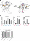
Figure 5. Determinants of receptor binding
(a) Virus-Overlay Protein Binding Assay (VOPBA) showing binding efficiency for different WE strains of LCMV. WE54 is known to exhibit high-affinity binding, while WE2.2 is known to exhibit low-affinity binding ,. (b) ELISA showing relative binding affinity for soluble DG. (c) Inhibition of viral infectivity in the presence of soluble αDG. Virus infection is shown as fluorescence focus units/mL (FFU/mL). (d) Subunit requirements for immunoprecipitation of αDG from cell lysates. Top panel, western blot for DG; bottom panel, western blot for LCMV GP1. For panels b and c error bars indicate s.d. (n=3 technical replicates or cell cultures, respectively). For panel d uncropped blots can be found in Supplementary Data Set 1.
Figure 6
Figure 6. The basic, lower loop face and dimeric interface are required for αDG binding
(a) Cartoon representation of the GP1 subunit of LCMV with residues analyzed shown as sticks. For clarity, the N-terminal strand of GP1 was removed from the representation. Mutations to residues in yellow did not produce virus with titers high enough to analyze further. Viruses bearing mutations to residues in blue (E109A, T111V, R185A, Q231A, and R233A) efficiently bound to αDG. Viruses bearing mutations to residues in magenta (H136R and R190A), failed to bind αDG. Residues previously known to confer high-affinity binding (S135 and L260) as well as Y155 (histidine in the crystallized strain) identified here are colored green. H136 at the dimeric interface is approximately 20Å from the 153, 155 and 190 cluster while L260 at the GP1-GP2 cleavage site is approximately 25Å from these three residues. (b) Inhibition of viral infectivity in the presence of soluble αDG. The y axis of each graph is the log decrease in virus infection upon preincubation with soluble αDG, as compared to pre-incubation with PBS. All point mutations were made in the H155Y background, and the H155Y single mutation is shown in each graph for comparison. (c) LCMV strains with both high- and low-αDG affinities infect BHK and LAMP1 knock-out cells with equal efficiency. Data shown represents the ratio of LCMV-positive LAMP1 knock-out cells to LCMV-positive BHK cells. Strains of LCMV that bind to αDG with high affinity are indicated by (+) and those that bind to αDG with low affinity are indicated by (–). For panels b and c error bars indicate the s.d. (n=3 cell cultures).
Figure 7
Figure 7. LCMV GP fit into tomographic reconstructions of LASV GPC on the viral surface
Single protomers of prefusion LCMV GPe dimer were docked into the tomographic reconstruction of Lassa virus (LASV) GPC spikes on the virion surface (EMD 3290 ). In this docking, all visualized glycans point outward and HR2 and membrane proximal regions orient to the membrane. The panel on the right shows the docked LCMV GP from the side; the panel on the left shows the GP from the top. Regions mapped to αDG binding are noted with a circle on each GP monomer for the cluster of residues 153, 155 and 190 (ranging from approximately 10-20Å from the top of the trimer); with arrows for residue 260 on each GP monomer, which is adjacent to the GP1-GP2 S1P cleavage site (approximately 45Å from the top of the trimer); and with stars for the dimeric interface residues 136 and 143.

Similar articles

Cited by

References

    1. Stephensen CB, et al. Prevalence of serum antibodies against lymphocytic choriomeningitis virus in selected populations from two U.S. cities. J Med Virol. 1992;38:27–31. - PubMed
    1. Childs JE, et al. Human-rodent contact and infection with lymphocytic choriomeningitis and Seoul viruses in an inner-city population. Am J Trop Med Hyg. 1991;44:117–21. - PubMed
    1. Ambrosio AM, Feuillade MR, Gamboa GS, Maiztegui JI. Prevalence of lymphocytic choriomeningitis virus infection in a human population of Argentina. Am J Trop Med Hyg. 1994;50:381–6. - PubMed
    1. Marrie TJ, Saron MF. Seroprevalence of lymphocytic choriomeningitis virus in Nova Scotia. Am J Trop Med Hyg. 1998;58:47–9. - PubMed
    1. Lledo L, Gegundez MI, Saz JV, Bahamontes N, Beltran M. Lymphocytic choriomeningitis virus infection in a province of Spain: analysis of sera from the general population and wild rodents. J Med Virol. 2003;70:273–5. - PubMed

Publication types

MeSH terms