Review
. 2016 May;36(3):303-16.
doi: 10.1111/opo.12283.

Infrared reflectance imaging in age-related macular degeneration

Affiliations
Review

Infrared reflectance imaging in age-related macular degeneration

Angelica Ly et al. Ophthalmic Physiol Opt. 2016 May.

Abstract

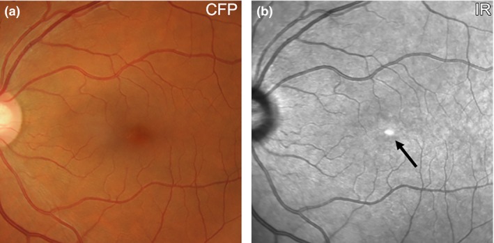
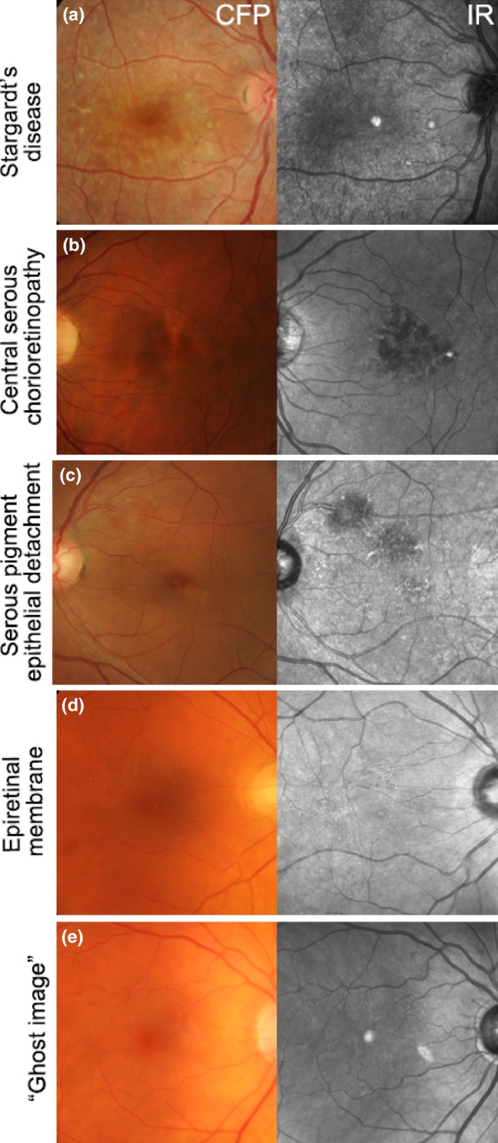
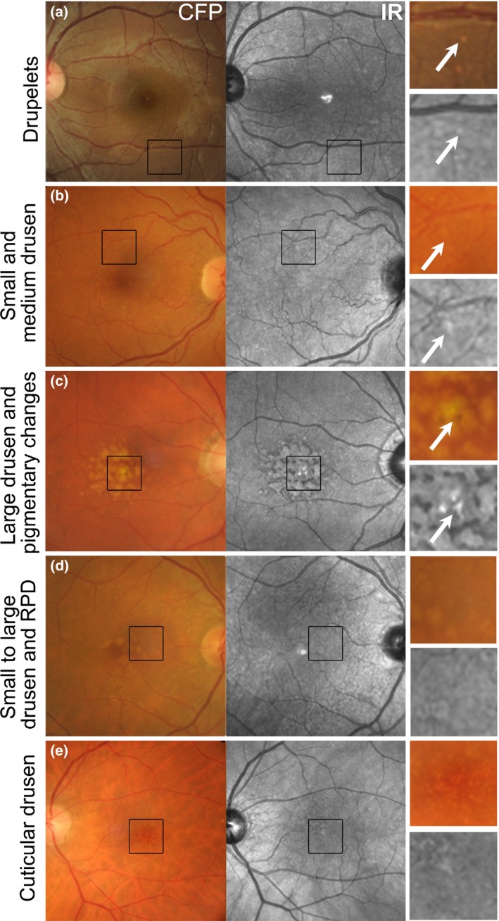
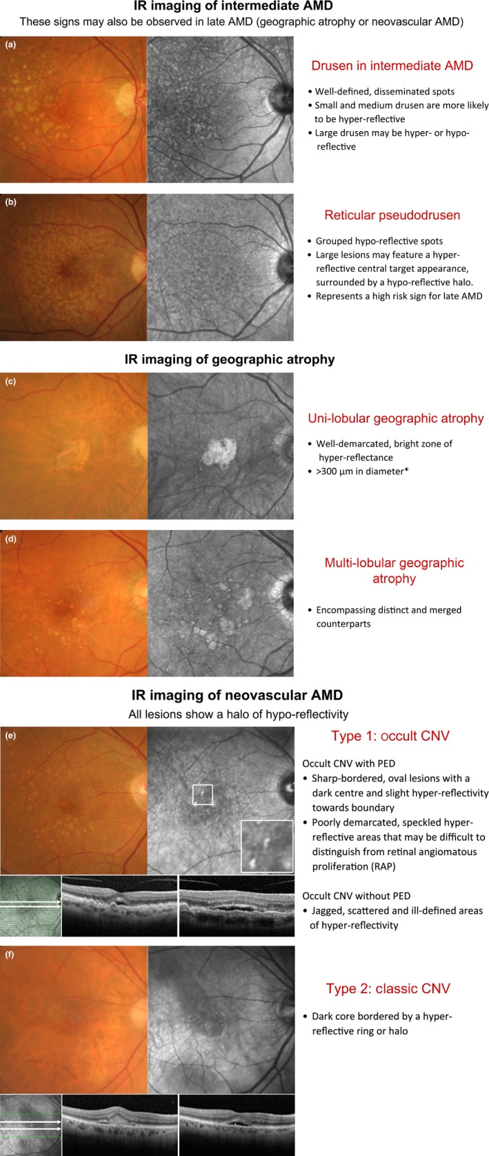
Purpose: The purpose of this article is to describe the appearance of age-related macular degeneration (AMD) phenotypes using infrared (IR) reflectance imaging. IR reflectance imaging of the retina has the potential to highlight specific sub-retinal features and pathology. However, its role in macular disease, specifically AMD, is often underestimated and requires clarification.

Recent findings: Recent advances in clinical methods, imaging and scientific knowledge may be integrated to improve the accuracy of disease stratification in AMD. In particular, IR imaging holds an underutilised sensitivity to detect reticular pseudodrusen, which have been repeatedly described as a high-risk sign for late AMD.

Summary: This article provides clinically relevant descriptions of AMD phenotypes using IR reflectance imaging. The findings are integrated with images from cases seen at the Centre for Eye Health. As primary eye-care providers assume a critical role in the detection, diagnosis and management of AMD, we also provide a chair-side reference to assist clinicians in interpreting IR images in AMD.

Keywords: age-related macular degeneration; chair-side reference; drusen; infrared; retina.


Figures

Figure 1
Figure 1
(a) Retinal photograph and (b) IR reflectance image of a 57 year old Caucasian female without any macular disease. The normal foveal ‘umbo’ appeared as a discrete, plaque‐like, hyper‐reflective spot in the IR image while the surrounding macula showed homogeneous reflectivity (arrow). Increased central IR signal may represent a reflection artefact or be attributable to the higher melanin content of foveal retinal pigment epithelium cells relative to more eccentric RPE. Choroidal and retinal vessels appear as dark shadows in the IR image. CFP, colour fundus photograph; IR, Infrared image.
Figure 2
Figure 2
Using IR imaging, macular lesions can appear hyper‐reflective as in (a) Stargardt's disease, or hypo‐reflective as in (b) Central serous chorioretinopathy, (c) Serous pigment epithelial detachment and (d) Epiretinal membrane. (e) Ghost image in an eye with a posterior chamber intraocular lens. CFP, colour fundus photograph; IR, Infrared image.
Figure 3
Figure 3
The appearance of drusen subtypes and pigment changes using IR imaging. (a) Drupelets (small drusen) present in a 29‐year‐old Caucasian female. (b) Early AMD characterised by small and medium sized drusen in a 69‐year‐old Caucasian female. The small and medium sized drusen in both cases appeared as well‐defined, disseminated spots of increased IR reflectivity. (c) Intermediate AMD in a 72‐year‐old Caucasian male with predominantly large drusen and central hyper‐pigmentary changes. The large drusen appear relatively well‐defined, mildly hyper‐reflective in comparison to the surrounds and are surrounded by hypo‐reflectivity in the IR image. Pigmentary changes appeared discrete and showed more intense hyper‐reflectivity than the drusen. (d) Intermediate AMD in a 73‐year‐old Caucasian female. The retinal photograph shows drusen ranging in size from small to large, predominantly affecting the central macula associated with variations in the greyscale tone of the IR image. Reticular pseudodrusen are also present and appear as grouped, hypo‐reflective spots most noticeable at the superior macula using the IR reflectance image. (e) Left eye images of a 65‐year‐old Caucasian female with cuticular drusen. The cuticular drusen appear punctate, hypo‐ and hyper‐reflective, and less distinct using IR imaging than in the CFP. CFP, colour fundus photograph; IR, Infrared image; RPD, reticular pseudodrusen.
Figure 4
Figure 4
Double‐sided chair side reference chart designed to aid in the differential diagnosis of AMD phenotypes using IR imaging: (a) Intermediate AMD and scattered drusen of various sizes. The small and medium sized drusen (<125 μm) were predominantly hyper‐reflective, while the effect of large drusen (≥125 μm) was more inconsistent (both hyper‐ and hypo‐reflective). (b) Reticular pseudodrusen (RPD), which typically present as a reticular pattern of small, yellow‐white, round lesions using colour retinal photography and as groups of hypo‐reflective lesions using IR imaging. Larger lesions may be accompanied by a halo‐like appearance, also known as a ‘target’ aspect, with increased IR centrally surrounded by a halo of decreased reflectivity. (c,d) Geographic atrophy secondary to AMD; the atrophic regions appear as well‐demarcated, bright zones of hyper‐reflectivity, which relates to the absence of RPE. The presentation may be (c) uni‐lobular involving the fovea or (d) multi‐lobular, extrafoveal and/or associated with drusen of variable sizes. Note that the atrophic areas may be more apparent using IR imaging than retinal photography. (e,f) Neovascular AMD; the IR images reveal (e) sharp‐bordered, oval lesions and speckled hyper‐reflectivity that suggest occult CNV, and (f) a dark core bordered by a hyper‐reflective ring or halo consistent with the typical appearance in classic CNV. Cases were chosen based on the consistency of the IR image to previous descriptions provided in the literature. *A size criterion of 300 μm was adopted by Xu et al.38 Historical characterisations of atrophic lesions in AMD 35 applied a 175 μm cut off to retinal photography observing the difficulties in detecting choroidal vessels and edges of atrophic areas in smaller regions.

References

    1. Bourne RR, Stevens GA, White RA et al Causes of vision loss worldwide, 1990–2010: a systematic analysis. Lancet Glob Health 2013; 1: e339–e349. - PubMed
    1. Wong WL, Su X, Li X et al Global prevalence of age‐related macular degeneration and disease burden projection for 2020 and 2040: a systematic review and meta‐analysis. Lancet Glob Health 2014; 2: e106–e116. - PubMed
    1. Chong GT & Lee RK. Glaucoma versus red disease: imaging and glaucoma diagnosis. Curr Opin Ophthalmol 2012; 23: 79–88. - PubMed
    1. Pecen PE & Kaiser PK. Current phase 1/2 research for neovascular age‐related macular degeneration. Curr Opin Ophthalmol 2015; 26: 188–193. - PubMed
    1. Age‐Related Eye Disease Study Research G . A randomized, placebo‐controlled, clinical trial of high‐dose supplementation with vitamins C and E, beta carotene, and zinc for age‐related macular degeneration and vision loss: AREDS report no. 8. Arch Ophthalmol 2001; 119: 1417–1436. - PMC - PubMed

Publication types