. 2016 May 9;26(9):1127-37.
doi: 10.1016/j.cub.2016.03.055. Epub 2016 Apr 21.

CDK1 Prevents Unscheduled PLK4-STIL Complex Assembly in Centriole Biogenesis

Affiliations

CDK1 Prevents Unscheduled PLK4-STIL Complex Assembly in Centriole Biogenesis

Sihem Zitouni et al. Curr Biol. .

Abstract

Centrioles are essential for the assembly of both centrosomes and cilia. Centriole biogenesis occurs once and only once per cell cycle and is temporally coordinated with cell-cycle progression, ensuring the formation of the right number of centrioles at the right time. The formation of new daughter centrioles is guided by a pre-existing, mother centriole. The proximity between mother and daughter centrioles was proposed to restrict new centriole formation until they separate beyond a critical distance. Paradoxically, mother and daughter centrioles overcome this distance in early mitosis, at a time when triggers for centriole biogenesis Polo-like kinase 4 (PLK4) and its substrate STIL are abundant. Here we show that in mitosis, the mitotic kinase CDK1-CyclinB binds STIL and prevents formation of the PLK4-STIL complex and STIL phosphorylation by PLK4, thus inhibiting untimely onset of centriole biogenesis. After CDK1-CyclinB inactivation upon mitotic exit, PLK4 can bind and phosphorylate STIL in G1, allowing pro-centriole assembly in the subsequent S phase. Our work shows that complementary mechanisms, such as mother-daughter centriole proximity and CDK1-CyclinB interaction with centriolar components, ensure that centriole biogenesis occurs once and only once per cell cycle, raising parallels to the cell-cycle regulation of DNA replication and centromere formation.

Keywords: CDK; PLK4; STIL; centriole duplication; centrosome; licensing; mitosis.


Conflict of interest statement

Competing interest statement

The authors declare no competing interests.

Figures

Figure 1
Figure 1. PLX4 induces de novo aster formation early after M-phase (CSF) release in Xenopus egg extracts
A. Cell cycle profiling of Xenopus M-phase extracts released with Ca2+. Western blots (WB) show the kinetics of Cdc25 downshift and CyclinB2 degradation leading to reduced CDK1 activity. B. GFP-PLX4AS-induces MTOC formation in M-phase released extracts. CSF extract was incubated with 0.65 μM GFP-PLX4AS, (green) and TRITC-tubulin (red). See also Figure S1, S2C for the generation and characterization of Shokat alleles. (See also Movie S1 and S2) C. Cell cycle-dependent effect of PLX4. rPLX4 was added at different time points: (i) 10 mins before release (add Ca2+), (ii) concomitantly with release, and (iii) 20 mins after release, in order to check when PLK4 activity is needed to form MTOCs (TRITC-tubulin). D. Inhibition of PLX4AS using 1 NA-PP1 at different stages. PLX4AS was added 10 mins before Ca2+ and was inhibited by 1NA-PP1 in extracts at different time points: (iv) at time zero (v) with Ca2+, and (vi) 20 mins after Ca2+. Panels of MTOCs formed in the extract under each condition, stained with TRITC-tubulin, are shown. Asters were counted in 10 images for each condition, and normalized to the number of asters observed in control extracts ((i) in C and PLX4AS in D) (n=3, Bars represent average +/SD).
Figure 2
Figure 2. PLK4 activity is needed at M-phase exit/G1 for centriole duplication in human cells
A–C. PLK4 activity was inhibited at different cell cycle times by 1NA-PP1 (PLK4AS) (B) or centrinone (C), and centrioles were counted in the subsequent mitosis. A. Experimental scheme and summary of results from B–C. B. 1NA-PP1 was added to cells expressing PLK4AS and depleted of endogenous PLK4 at 0 hrs (positive control, (+)), 6 (G2), 8 (M), 10 (M-exit/G1) and 20 hrs (S-phase) following S-phase block release. Cells were fixed in the immediate subsequent mitosis, and centrioles counted (n=3, 100 cells/condition, mean +/− SEM. ** = p<0.01). Cell cycle profiles were obtained by flow cytometry. Percentage of S-phase cells at time of inhibition were monitored by EdU staining (See also Figure S2F–I for controls). C. Centrinone was added at the indicated cell cycle stages and centrioles were counted in the subsequent mitosis. (n=3, 100 cells/condition. mean +/− SEM. ** = p<0.01). Representative immunofluorescence images of mitotic cells stained as indicated, following inhibition of PLK4 by centrinone. The number of centrioles found with the highest frequency upon inhibition is shown (average of 3 experiments). See Figure S2J for detailed quantitation.
Figure 3
Figure 3. The PLK4-STIL complex forms only upon M-exit in Xenopus extracts and human cells
A. PLX4 binds STIL only after mitotic-exit in Xenopus extracts. (See also Figure S3A–D). rPLX4 was supplemented to extracts at the indicated stages, and subsequently immunoprecipitated (PLX4-IP). Samples were analyzed by WB, and probed with the indicated antibodies. (*) refers to an interphase extract (see methods). B. PLX4 is likely to phosphorylate STIL upon M-exit in Xenopus extracts. Released-extracts were supplemented with rPLX4. STIL phosphorylation (note the mobility shift) and levels of CyclinB2 were analyzed by WB. C. STIL peaks in mitosis in human cells. FLAG-PLK4 over-expressing HeLa cells were synchronized in mitosis by monastrol, and released. Samples were collected at the indicated times. D. The PLK4-STIL complex forms in mid-G1. FLAG-PLK4 was immuno-precipitated from cell lysates at the indicated stages. Samples were probed with the indicated antibodies. Cell cycle profiles were obtained by flow cytometry combined with BrdU-FITC labeling of cells in S phase. See Figure S3E for more details. E. STIL recruitment to the centrosome during mid-G1 phase is dependent on PLK4 activity. Presence of STIL at the centrosome was quantified by immunofluorescence using anti-STIL in mitotic or G1 cells obtained after mitotic shake-off. DMSO (control) or centrinone (PLK4 inhibitor) was added 1hr prior to fixation. The absence of S-phase cells in G1 was corroborated by EdU incorporation. (n=3, 100 cells/condition, mean +/− SEM). See also Figure S3F, S3G, S4A and S4B for more details on these experiments.
Figure 4
Figure 4. PLK4-STIL complex formation is inhibited by CDK1. A. Inhibiting CDK1 allows PLK4-STIL complex assembly
STIL was immunoprecipitated from extracts treated as indicated. Samples were analyzed by WB, probed with the indicated antibodies. Fold increase over control in signal intensity was quantified for each condition. Note that endogenous PLX4 is undetectable. *Addition of RO-3306, a CDK1 inhibitor, releases the extract into interphase. B. Phosphorylation of STIL in CSF/M-phase arrested extracts treated with RO-3306. M-phase extracts were treated as indicated. STIL phosphorylation and CyclinB2 were analyzed by WB. C. PLX4 induces asters in M-phase extracts released with RO-3306. Confocal images of M-phase extracts supplemented with rPLX4 and 100 μM RO-3306, in the presence of TRITC-labeled tubulin. D. CDK1/CyclinB activity is inhibitory for the formation of the PLK4-STIL complex. Nocodazole arrested cells in mitosis were treated as indicated. STIL was immunoprecipitated and samples were probed with the indicated antibodies. Note that under centrinone treatment, PLK4 accumulates [30]. Cell cycle profiles were obtained by flow cytometry; see Figure S4C for details.
Figure 5
Figure 5. CDK1/CyclinB phosphorylates STIL outside of the PLK4 interacting domain in human cells
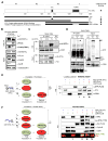
A. Schematic representation of HA- and GST-tagged STIL constructs. The evolutionary conserved coiled-coil (CC; PLK4-binding) and STAN (PLK4-phosphorylated and SAS6 binding) domains are indicated. Constructs were used for phosphorylation assays. The results are summarized on the right. B. CDK1/ Cyclin B can phosphorylate STIL on its N-terminus. Autoradiography of an In vitro kinase assay using IP control, or HA-STIL (FL, ΔCC, N and N3C), CDK1/Cyclin B and ([γ-32P]-ATP). Black arrows show phosphorylated fragments. White arrow indicates the expected position of the non-phosphorylated N3C fragment (n.s.=non-specific). Note that all fragments, but not the N3C, are phosphorylated by CDK1/CyclinB. C. STIL-N3C, the domain that interacts with PLK4 and SAS6, is not phosphorylated by CDK1/CyclinB. Incorporation of [γ-32P] on STIL-N3C incubated with GFP-PLX4 or CDK1/CyclinB was visualized by autoradiography. D. CDK1/CyclinB does not phosphorylate STIL on the site phosphorylated by PLK4 (S1116). Recombinant GST-STIL (FL) was incubated with rPLX4 or CDK1/CyclinB. Samples were analyzed by WB using anti-pS1116-STIL (which recognizes a PLK4-specific phosphorylation [8]), anti-His and anti-CDK1 to monitor loaded proteins.
Figure 6
Figure 6. CDK1/Cyclin B binds STIL on its CC domain and competes with PLK4 binding
A. Representation of HA-, GST- and GFP-tagged STIL constructs and summary of results. The evolutionary conserved coiled-coil (CC; PLK4 binding) and STAN (PLK4 phosphorylated and SAS6 binding) domains are indicated. B. CDK1/CyclinB and PLK4 bind STIL at different stages. rGST-STIL and rPLX4 were added to CSF extracts which were kept arrested or released to interphase with Ca2+. GST-STIL was immunoprecipitated and samples were analyzed by WB. See Figure S5D and S5E. C. The STIL-CC motif is necessary for CDK1/CyclinB binding. HEK293T cells were transfected with the indicated plasmids and CDK1 was immunoprecipitated. Samples were visualized by WB using the indicated antibodies. D. The STIL-CC motif is sufficient for CDK1/CyclinB binding. CDK1 was immunoprecipitated from HEK293T cells expressing GFP, GFP-CC or GFP-FL-STIL. Co-immunoprecipitates were probed with the indicated antibodies (n.s.= non-specific; cross reaction of the mouse anti-GFP antibody with rabbit IgGs used to IP CDK1). E. CDK1 binding to STIL prevents subsequent PLX4 binding. GST-N3CΔSTAN was incubated as indicated in the scheme, pulled-down, and its co-precipitated proteins were analyzed by WB. See Figure S6A and S6B for quantification (n=3) and titration experiments. F. CDK1 binding of STIL prevents its phosphorylation by PLX4. GST-STIL (FL) was incubated as shown in the scheme. STIL phosphorylation by PLX4 in the STAN motif was analyzed by WB using the indicated antibodies. See Figure S6C for more details on this experiment.

Similar articles

Cited by

References

    1. Bettencourt-Dias M, Glover DM. Centrosome biogenesis and function: centrosomics brings new understanding. Nat Rev Mol Cell Biol. 2007;8:451–463. - PubMed
    1. Khodjakov A, Rieder CL, Sluder G, Cassels G, Sibon O, Wang C-L. De novo formation of centrosomes in vertebrate cells arrested during S phase. J Cell Biol. 2002;158:1171–81. - PMC - PubMed
    1. Zitouni S, Nabais C, Jana SC, Guerrero A, Bettencourt-Dias M. Polo-like kinases: structural variations lead to multiple functions. Nat Rev Mol Cell Biol. 2014;15:433–452. - PubMed
    1. Bettencourt-Dias M, Rodrigues-Martins A, Carpenter L, Riparbelli M, Lehmann L, Gatt MK, Carmo N, Balloux F, Callaini G, Glover DM. SAK/PLK4 is required for centriole duplication and flagella development. Curr Biol. 2005;15:2199–2207. - PubMed
    1. Habedanck R, Stierhof Y-D, Wilkinson CJ, Nigg EA. The Polo kinase Plk4 functions in centriole duplication. Nat Cell Biol. 2005;7:1140–1146. - PubMed

MeSH terms

Substances

LinkOut - more resources