. 2016 Aug;29(8):930-40.
doi: 10.1111/tri.12788. Epub 2016 Jun 14.

Regulatory T cell expressed MyD88 is critical for prolongation of allograft survival

Affiliations

Regulatory T cell expressed MyD88 is critical for prolongation of allograft survival

Christopher M Borges et al. Transpl Int. 2016 Aug.

Abstract

MyD88 signaling directly promotes T-cell survival and is required for optimal T-cell responses to pathogens. To examine the role of T-cell-intrinsic MyD88 signals in transplantation, we studied mice with targeted T-cell-specific MyD88 deletion. Contrary to expectations, we found that these mice were relatively resistant to prolongation of graft survival with anti-CD154 plus rapamycin in a class II-mismatched system. To specifically examine the role of MyD88 in Tregs, we created a Treg-specific MyD88-deficient mouse. Transplant studies in these animals replicated the findings observed with a global T-cell MyD88 knockout. Surprisingly, given the role of MyD88 in conventional T-cell survival, we found no defect in the survival of MyD88-deficient Tregs in vitro or in the transplant recipients and also observed intact cell homing and expression of Treg effector molecules. MyD88-deficient Tregs also fail to protect allogeneic bone marrow transplant recipients from chronic graft-versus-host disease, confirming the observations of defective regulation seen in a solid organ transplant system. Together, our data define MyD88 as having a divergent requirement for cell survival in non-Tregs and Tregs, and a yet-to-be defined survival-independent requirement for Treg function during the response to alloantigen.

Keywords: T cells; Treg; inflammation; transplantation.


Figures

Figure 1
Figure 1
A. bm12 skin allografts were transplanted onto WT or MyD88-ΔT recipients. Mice received either no treatment (● and ∎, respectively), or i.p. injection of 0.25 mg αnti-CD154 (Clone: MR1, administered on Days 0, 2 and 4) and i.p. injection of 1 mg/kg rapamycin (administered on Days 0, 2, 4, 6, 8, 10 and 12) (❍ and ◻, respectively). Data are pooled from 2 independent experiments. MST: 13, 11, 90, 53 days (●, ∎, ❍, ◻) respectively. B. bm12 cardiac allografts were transplanted into WT or MyD88-ΔT recipients. Mice received either no treatment (●and ∎, respectively), or i.v. injection of 0.5×10⁶ sorted WT Tregs (isolated from FoxP3GFP mice 7 days prior to transplant (◻). Data are pooled from 2 independent experiments. MST: undefined, 34 days, and undefined (●,∎,◻) respectively. *, p<0.05. **, p<0.01.
Figure 2
Figure 2
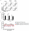
A. Representative flow cytometry plots of sort-purified CD4+CD25+ Tregs or CD4+CD25+CD62L+CD44 naïve, non- Tregs from WT (white bars) or MyD88-ΔT (black bars) mice stained with annexin V and 7-AAD after 72 hours of in vitro culture with anti-CD3 and anti-CD28, with and without 10 ng/mL IL-2. Numbers in lower-left quadrant indicate frequency of cells within that quadrant. B. Quantification of data from 5 independent experiments of A. C. Female MyD88+/+ Foxp3+/Cre-YFP (Foxp3-Cre-YFP het) MyD88fl/+ FoxP3+/Cre-YFP (MyD88fl/+ Foxp3-Cre-YFP het) and MyD88fl/flxFoxp3+/Cre-YFP (MyD88fl/fl Foxp3-Cre-YFP het) mice were bled bi-weekly starting at 6 weeks of age for 26 weeks. Percent YFP positive cells were assessed among total Foxp3+CD4+ antibody stained cells. Data are pooled from a minimum of 2 independent experiments. Error bars display standard deviation. **, p<0.01.
Figure 3
Figure 3
WT (black lines and black bars, minimum n=10 mice) and MyD88-ΔTreg (red lines and red bars, minimum n=11 mice) CD4+Foxp3+ cells were isolated from pooled spleen and peripheral lymph nodes. Cells were analyzed directly ex vivo, or stimulated with anti-CD3 and anti-CD28 for 20 hours. At each time point, A. CD39 and CD73, B. surface CTLA-4, C. intracellular CTLA-4, D. Lag-3 and E. Granzyme B were assessed. Shaded gray area indicates appropriate isotype control for indicated stain. Representative flow cytometry plots shown from a minimum of 3 independent experiments. F. In vitro suppression assay. Quantification of percent proliferation of WT naïve T cells co-cultured with irradiated APCs and indicated ratios of WT (black bars) or MyD88-ΔTreg (red bars) (normalized to proliferation of WT naïve cells without Treg). Data are pooled from 5 independent experiments. Error bars display standard deviation.
Figure 4
Figure 4
bm12 skin allografts were transplanted onto WT (n=14 recipients, 7 male Cre-hemizygous mice, 7 female homozygous mice). MyD88fl/flFoxp3+/Cre heterozygous (n=5 female Cre heterozygous recipients) or MyD88-ΔTreg (n=12 recipients, 8 male Cre-hemizygous mice, 4 female Cre-homozygous mice) treated with CoB as in Figure 1.1 (❍, ◇ and ◻, respectively). Data are pooled from 3 independent experiments. MST: undefined, 37 and 59 days (❍, ◇, ◻) respectively. ***, p<0.001.
Figure 5
Figure 5
A. bm12 skin grafts were harvested from WT (white bars, n≥5) or MyD88-ΔTreg (black bars, n≥3) recipient mice, and frequency of Foxp3+ CD4+ cells was assessed by flow cytometry at each indicated time point post transplant. B & C. Intragraft Foxp3+ (panel B) and Foxp3 (panel C) cellularity was calculated using the number of CD45+ cells isolated from WT (white bars, n=5) or MyD88-ΔTreg (black bars, n=6) recipient mice with a bead based flow cytometric counting assay; data are representative of 2 independent experiments. D. Skin grafts were harvested from WT (white bars, n=5) or MyD88-ΔTreg (black bars, n=6) recipient mice, digested and stained for Foxp3 and with Live/Dead Aqua. Frequency of Live/Dead Aqua positive cells are indicated, data are representative of 2 independent experiments. E. Fluorescent imaging of skin grafts from WT or MyD88-ΔTreg recipient mice. Sections were imaged at 10x and 40x magnification. White box on 10x magnification images indicate area imaged with 40x objective. Data are representative of grafts harvested from n=6 WT mice and n=8 MyD88-ΔTreg mice. F. Frequency of Annexin V- 7-AAD- WT (white bars) or MyD88-ΔTreg (black bars) Treg after activation and culture with anti-CD3, anti-CD28 and IL-2 with indicated concentrations of αnti-CD154 and/or rapamycin for 72 hours. Quantification of data from a minimum of 3 independent experiments. Error bars display standard deviation. *, p<0.05.
Figure 6
Figure 6
A. Mouse weight and B. survival curve following induction of cGVHD in B10.BR mice following B6 bone marrow transplant (●) and B6 bone marrow transplant plus adoptive transfer of either WT (❍) or MyD88-ΔTreg (◻) splenocytes. C. Airway resistance and D. compliance measured on Day 28 post BMT from recipients of B10.BR bone marrow alone (gray bars), B10.BR bone marrow plus WT splenocytes (white bars) or B10.BR bone marrow plus MyD88-ΔTreg splenocytes (black bars). Error bars display standard error of the mean. *, p<0.05. **, p<0.01. ***, p<0.001.

References

    1. Medzhitov R, Preston-Hurlburt P, Kopp E, Stadlen A, Chen C, Ghosh S, et al. MyD88 is an adaptor protein in the hToll/IL-1 receptor family signaling pathways. Mol Cell. 1998;2(2):253–8. - PubMed
    1. Casanova JL, Abel L, Quintana-Murci L. Human TLRs and IL-1Rs in host defense: natural insights from evolutionary, epidemiological, and clinical genetics. Annu Rev Immunol. 2011;29:447–91. - PubMed
    1. Gelman AE, LaRosa DF, Zhang J, Walsh PT, Choi Y, Sunyer JO, et al. The adaptor molecule MyD88 activates PI-3 kinase signaling in CD4+ T cells and enables CpG oligodeoxynucleotide-mediated costimulation. Immunity. 2006;25(5):783–93. - PMC - PubMed
    1. Gelman AE, Zhang J, Choi Y, Turka LA. Toll-like receptor ligands directly promote activated CD4+ T cell survival. J Immunol. 2004;172(10):6065–73. - PMC - PubMed
    1. LaRosa DF, Stumhofer JS, Gelman AE, Rahman AH, Taylor DK, Hunter CA, et al. T cell expression of MyD88 is required for resistance to Toxoplasma gondii. Proc Natl Acad Sci U S A. 2008;105(10):3855–60. - PMC - PubMed

MeSH terms

LinkOut - more resources