Practical one-dimensional measurements of age-related brain atrophy are validated by 3-dimensional values and clinical outcomes: a retrospective study
- PMID: 27113039
- PMCID: PMC4845392
- DOI: 10.1186/s12880-016-0136-x
Practical one-dimensional measurements of age-related brain atrophy are validated by 3-dimensional values and clinical outcomes: a retrospective study
Abstract
Background: Age-related brain atrophy has been represented by simple 1-dimensional (1-D) measurements on computed tomography (CT) for several decades and, more recently, with 3-dimensional (3-D) analysis, using brain volume (BV) and cerebrospinal fluid volume (CSFV). We aimed to show that simple 1-D measurements would be associated with 3-D values of age-related atrophy and that they would be related to post-traumatic intracranial hemorrhage (ICH).
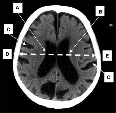
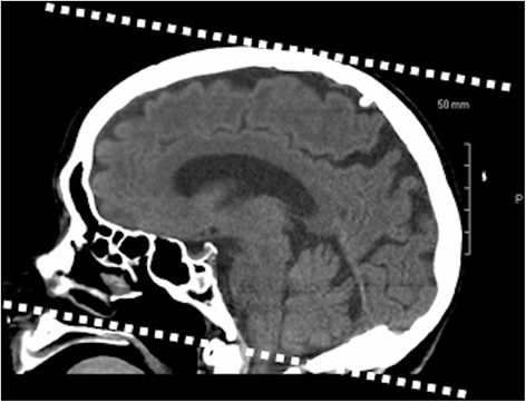
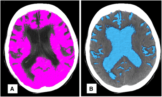
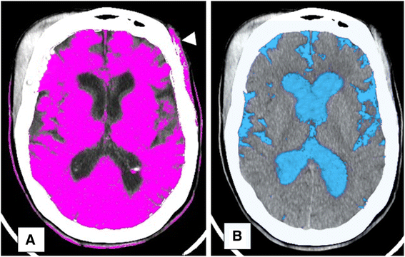
Methods: Patients ≥60 years with head trauma were classified with central atrophy (lateral ventricular body width >30 mm) and/or cortical atrophy (sulcus width ≥2.5 mm). Composite atrophy was the presence of central or cortical atrophy. BV and CSFV were computed using a Siemens Syngo workstation (VE60A).
Results: Of 177 patients, traits were age 78.3 ± 10, ICH 32.2%, central atrophy 39.5%, cortical atrophy 31.1%, composite atrophy 49.2%, BV 1,156 ± 198 mL, and CSFV 102.5 ± 63 mL. CSFV was greater with central atrophy (134.4 mL), than without (81.7 mL, p < 0.001). BV was lower with cortical atrophy (1,034 mL), than without (1,211 mL; p < 0.001). BV was lower with composite atrophy (1,103 mL), than without (1,208 mL; p < 0.001). CSFV was greater with composite atrophy (129.1 mL), than without (76.8 mL, p < 0.001). CSFV÷BV was greater with composite atrophy (12.3%), than without (6.7%, p < 0.001). Age was greater with composite atrophy (80.4 years), than without (76.3, p = 0.006). Age had an inverse correlation with BV (p < 0.001) and a direct correlation with CSFV (p = 0.0002) and CSFV÷BV (p < 0.001). ICH was greater with composite atrophy (49.4%), than without (15.6%; p < 0.001; odds ratio = 5.3). BV was lower with ICH (1,089 mL), than without (1,188 mL; p = 0.002). CSFV÷BV was greater with ICH (11.1 %), than without (8.7%, p = 0.02). ICH was independently associated with central atrophy (p = 0.001) and cortical atrophy (p = 0.003).
Conclusions: Simple 1-D measurements of age-related brain atrophy are associated with 3-D values. Clinical validity of these methods is also supported by their association with post-injury ICH. Intracranial 3-D software is not available on many CT scanners and can be cumbersome, when available. Simple 1-D measurements, using the study methodology, are a practical method to objectify the presence of age-related brain atrophy.
Keywords: Brain atrophy; CT imaging; Traumatic intracranial hemorrhage.
Figures


Similar articles
-
Impact of brain volume and intracranial cerebrospinal fluid volume on the clinical outcome in endovascularly treated stroke patients.J Stroke Cerebrovasc Dis. 2020 Jul;29(7):104831. doi: 10.1016/j.jstrokecerebrovasdis.2020.104831. Epub 2020 May 11. J Stroke Cerebrovasc Dis. 2020. PMID: 32404285
-
Measuring Global Brain Atrophy with the Brain Volume/Cerebrospinal Fluid Index: Normative Values, Cut-Offs and Clinical Associations.Neurodegener Dis. 2016;16(1-2):77-86. doi: 10.1159/000442443. Epub 2015 Dec 19. Neurodegener Dis. 2016. PMID: 26726737
-
Are routine repeat imaging and intensive care unit admission necessary in mild traumatic brain injury?J Neurosurg. 2012 Mar;116(3):549-57. doi: 10.3171/2011.11.JNS111092. Epub 2011 Dec 23. J Neurosurg. 2012. PMID: 22196096
-
Loss of entorhinal cortex and hippocampal volumes compared to whole brain volume in normal aging: the SMART-Medea study.Psychiatry Res. 2012 Jul 30;203(1):31-7. doi: 10.1016/j.pscychresns.2011.12.002. Epub 2012 Aug 19. Psychiatry Res. 2012. PMID: 22910574
-
[Imaging in geriatric medicine].Nihon Ronen Igakkai Zasshi. 1992 Jan;29(1):17-23. doi: 10.3143/geriatrics.29.17. Nihon Ronen Igakkai Zasshi. 1992. PMID: 1560604 Review. Japanese. No abstract available.
Cited by
-
Novel multi-linear quantitative brain volume formula for manual radiological evaluation of brain atrophy.Eur J Radiol Open. 2020 Nov 13;7:100281. doi: 10.1016/j.ejro.2020.100281. eCollection 2020. Eur J Radiol Open. 2020. PMID: 33241090 Free PMC article.
-
Fully Automatic Classification of Brain Atrophy on NCCT Images in Cerebral Small Vessel Disease: A Pilot Study Using Deep Learning Models.Front Neurol. 2022 Mar 24;13:846348. doi: 10.3389/fneur.2022.846348. eCollection 2022. Front Neurol. 2022. PMID: 35401411 Free PMC article.
-
High Blood Lead Levels: An Increased Risk for Development of Brain Hyperintensities among Type 2 Diabetes Mellitus Patients.Biol Trace Elem Res. 2021 Jun;199(6):2149-2157. doi: 10.1007/s12011-020-02359-6. Epub 2020 Aug 31. Biol Trace Elem Res. 2021. PMID: 32865724
References
-
- Steiner I, Gomori JM, Melamed E. Progressive brain atrophy during normal aging in man: a quantitative computerized tomography study. Isr J Med Sci. 1985;21(3):279–282. - PubMed
MeSH terms
LinkOut - more resources
Full Text Sources
Other Literature Sources
Medical

