. 2016 Apr 26:6:25013.
doi: 10.1038/srep25013.

p58(IPK) suppresses NLRP3 inflammasome activation and IL-1β production via inhibition of PKR in macrophages

Affiliations

p58(IPK) suppresses NLRP3 inflammasome activation and IL-1β production via inhibition of PKR in macrophages

Evgenii Boriushkin et al. Sci Rep. .

Abstract

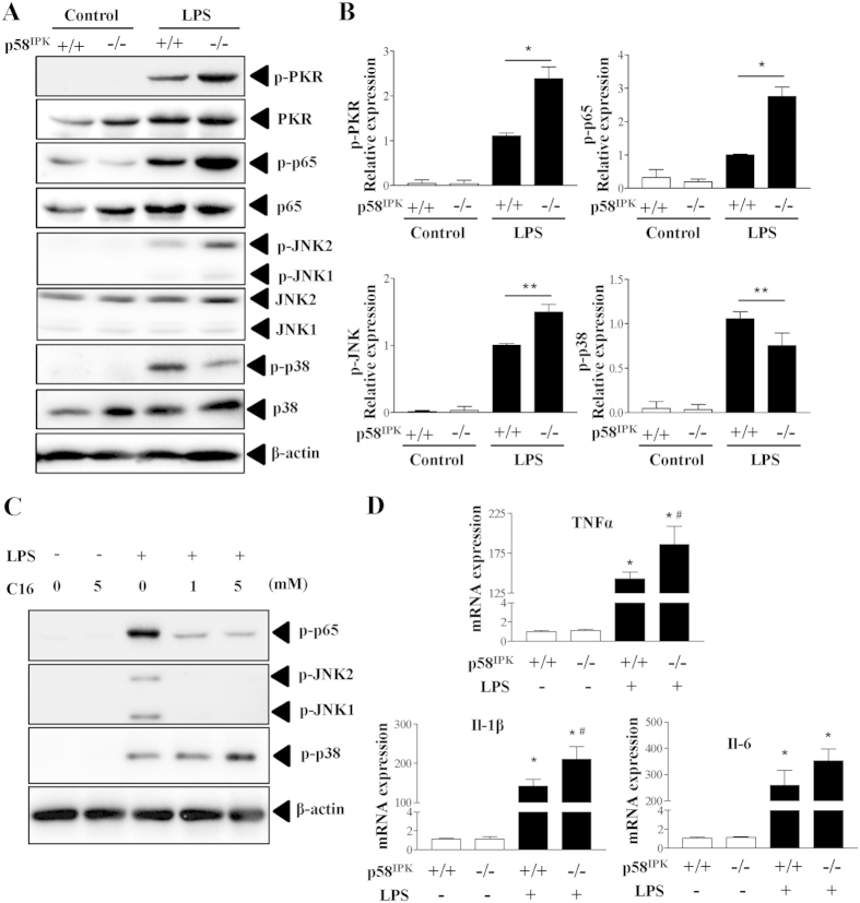
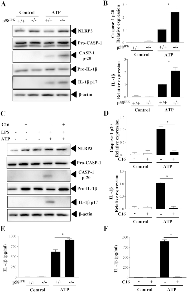
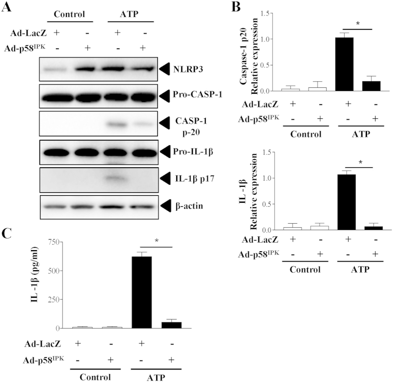
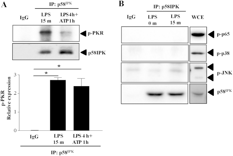
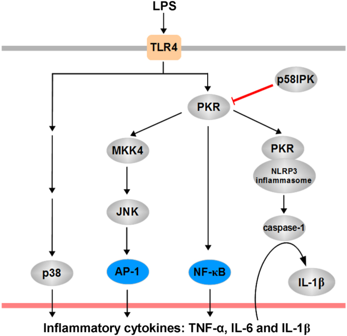
The NLRP3 inflammasome activation is a key signaling event for activation and secretion of pro-inflammatory cytokines such as IL-1β from macrophages. p58(IPK) is a molecular chaperone that regulates protein homeostasis through inhibiting eIF-2α kinases including double-stranded RNA-dependent protein kinase (PKR), which has been recently implicated in inflammasome activation. Herein we investigate the role of p58(IPK) in TLR4 signaling and inflammasome activation in macrophages. Primary bone marrow-derived macrophages (BMDM) was isolated from p58(IPK) knockout (KO) and wildtype (WT) mice and treated with lipopolysaccharide (LPS) and ATP to activate TLR4 signaling and stimulate inflammasome activation. Compared to WT macrophages, p58(IPK) deficient cells demonstrated significantly stronger activation of PKR, NF-κB, and JNK and higher expression of pro-inflammatory genes TNF-α and IL-1β. Coincidently, p58(IPK) deletion intensified NLRP3-inflammasome activation indicated by enhanced caspase 1 cleavage and increased IL-1β maturation and secretion. Pretreatment with specific PKR inhibitor or overexpression of p58(IPK) largely abolished the changes in inflammasome activation and IL-1β secretion in p58(IPK) null macrophages. Furthermore, immunoprecipitation assay confirmed the binding of p58(IPK) with PKR, but not other TLR4 downstream signaling molecules. Collectively, these results suggest a novel and crucial role of p58(IPK) in regulation of inflammasome activation and IL-1β secretion in macrophages.


Figures

Figure 1
Figure 1. p58IPK regulates LPS-induced activation of PKR, NF-κB and JNK MAPK pathways.
(A,B) BMDM isolated from WT (p58IPK+/+) or p58IPK knockout (p58IPK−/−) mice were cultured and treated with LPS for 15 min. Phosphorylation of PKR, NF-Κb p65, JNK, and p38 MAPK was evaluated by western blot analysis and quantified by densitometry. β-actin was used as a loading control. Data represent means ± SD from at least 3 independent experiments. *P < 0.05 and **P < 0.01. (C) BMDM from WT mice were pretreated with specific PKR inhibitor C-16 and then exposed to LPS or vehicle for 15 min. Whole cell lysates were subjected to western blot analysis. (D) BMDM from WT (p58IPK+/+) or p58IPK knockout (p58IPK−/−) mice were treated with LPS for 2 h. mRNA expressions of TNFα, IL-1β and IL-6 were determined by real-time qPCR and normalized by 18S. Data represent means ± SD of 3 independent experiments. *P < 0.05 vs. no LPS treatment, #p < 0.05 vs LPS-treated p58IPK+/+ cells.
Figure 2
Figure 2. p58IPK deficiency exacerbates NLRP3 inflammasome activation via PKR.
(A,B) BMDM from WT (p58IPK+/+) or p58IPK knockout (p58IPK−/−) mice were primed with LPS (500 ng/ml) for 4 h and then stimulated with ATP (5 mM) for 1 h. Protein levels of NLRP3, pro- and cleaved capase-1 and IL-1β were determined by western blot analysis and quantified by densitometry. Data represent means ± SD of 3 independent experiments. *P < 0.01. (C,D) BMDM from p58IPK knockout (p58IPK−/−) mice were pretreated with C16 and then exposed to LPS and ATP to induce inflammasome activation. Whole cell lysates were assessed by western blot analysis. Protein levels were quantified by densitometry and normalized by β-actin. Data represent means ± SD of at least 3 independent experiments. *P < 0.01. (E,F) Levels of IL-1β secreted into the medium of p58IPK+/+ and p58IPK−/− BMDM cultures were measured by ELISA. Data represent means ± SD of at least 3 independent experiments. *P < 0.01.
Figure 3
Figure 3. Overexpression of p58IPK reduces inflammasome activation and IL-1β secretion.
(A,B) BMDM from WT mice were transduced with Ad-p58IPK or Ad-LacZ at MOI of 50 for 24 h. Cells were then treated with LPS and ATP to induce inflammasome activation. Whole cell lysates were subjected to western blot analysis. Protein levels were quantified by densitometry and normalized by β-actin. Data represent means ± SD of 3 independent experiments. *P < 0.01. (C) Levels of IL-1β secreted into the medium of BMDM were measured by ELISA. Data represent means ± SD of 3 independent experiments. *P < 0.01.
Figure 4
Figure 4. p58IPK deficiency does not alter macrophage adhesion to endothelial cells.
BMDM from WT (p58IPK+/+) or p58IPK knockout (p58IPK−/−) mice were treated with LPS (250 ng/ml) or vehicle for 6 h. Cells were labeled with fluorescent dye PKH26 and then added to HUVECs for adhesion assay. (A) Representative images of showing BMDM (red) adhering to HUVECs. LPS treatment increased BMDM adhesion to endothelial cells. However, no difference was observed in LPS-treated p58IPK−/− macrophages compared to p58IPK+/+ controls. (B) Quantification of adhered BMDM to HUVECs.
Figure 5
Figure 5. Interaction between p58IPK and PKR in BMDM.
(A) Mouse BMDM were treated as indicated and harvested. Cell lysate was subjected to immunoprecipitation using antibody against p58IPK or non-immunized serum (IgG) as a negative control, followed by western blot analysis. Relative protein levels of PKR were determined by densitometry. Data represent means ± SD of 3 independent experiments. (B) No positive binding of p58IPK with NF-κB p65, p38 or JNK was revealed in BMDM treated with LPS for 15 min, but strong activation of each molecule was observed in the whole cell extract (WCE) from cells under the same treatment. The gels were run under the same experimental conditions, and full-length blots are presented in Supplementary data.
Figure 6
Figure 6. Schematic summary of the role of p58IPK in regulation PKR-dependent signaling and proinflammatory cytokine production.

References

    1. Takeuchi O. & Akira S. Pattern recognition receptors and inflammation. Cell 140, 805–820, 10.1016/j.cell.2010.01.022 (2010). - DOI - PubMed
    1. Schroder K. & Tschopp J. The inflammasomes. Cell 140, 821–832, 10.1016/j.cell.2010.01.040 (2010). - DOI - PubMed
    1. Federici S., Martini A. & Gattorno M. The central role of anti-IL-1 blockade in the treatment of monogenic and multifactorial autoinflammatory diseases. Front. Immunol. 4, 10.3389/fimmu.2013.00351 (2013). - DOI - PMC - PubMed
    1. Wen H. et al. Fatty acid-induced NLRP3-ASC inflammasome activation interferes with insulin signaling. Nat. Immunol. 12, 408–415, 10.1038/ni.2022 (2011). - DOI - PMC - PubMed
    1. Hewett S. J., Jackman N. A. & Claycomb R. J. Interleukin-1β in Central Nervous System Injury and Repair. Eur. J. Neurodegener. Dis. 1, 195–211 (2012). - PMC - PubMed

Publication types

MeSH terms