. 2016 Apr 26:7:11401.
doi: 10.1038/ncomms11401.

Line tension at lipid phase boundaries as driving force for HIV fusion peptide-mediated fusion

Affiliations

Line tension at lipid phase boundaries as driving force for HIV fusion peptide-mediated fusion

Sung-Tae Yang et al. Nat Commun. .

Abstract

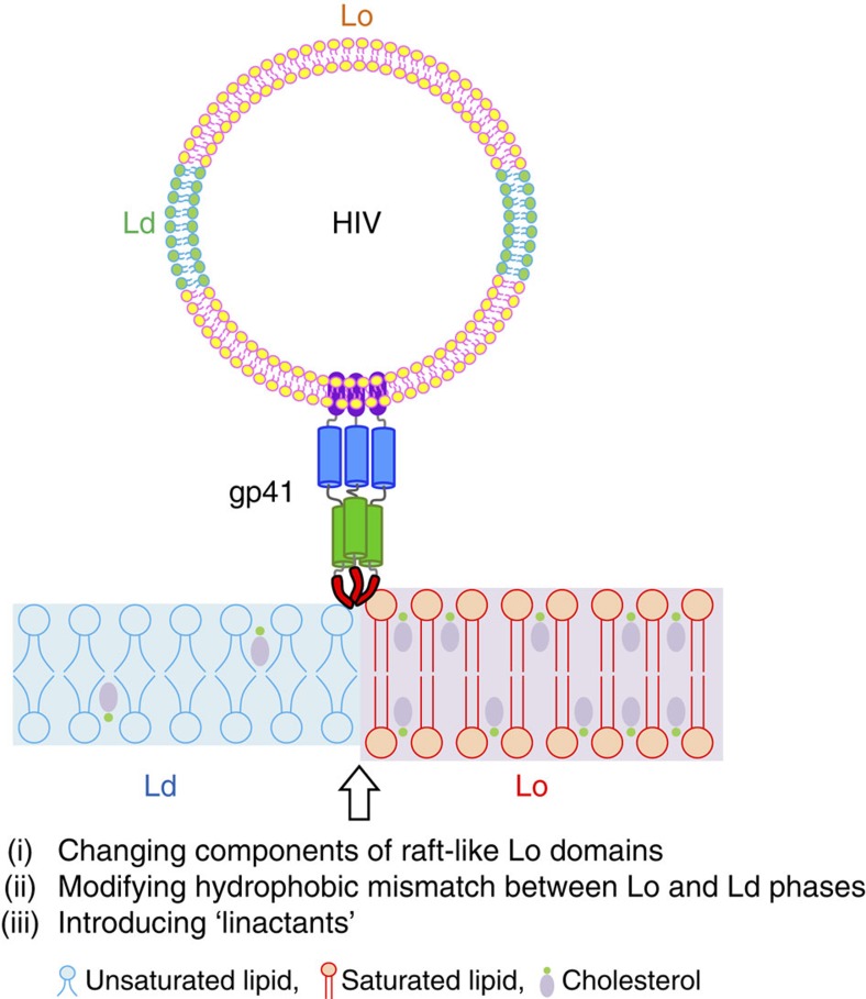
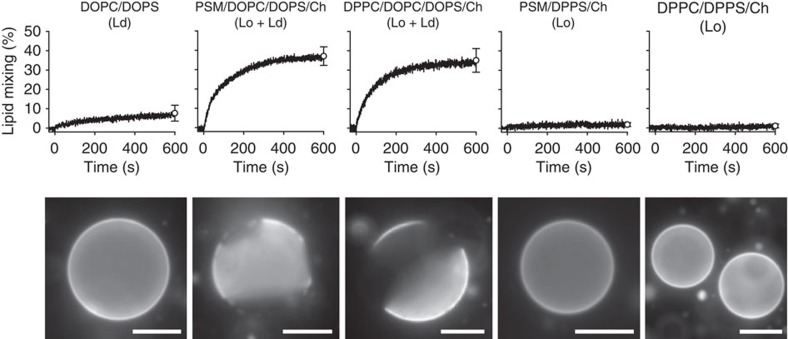
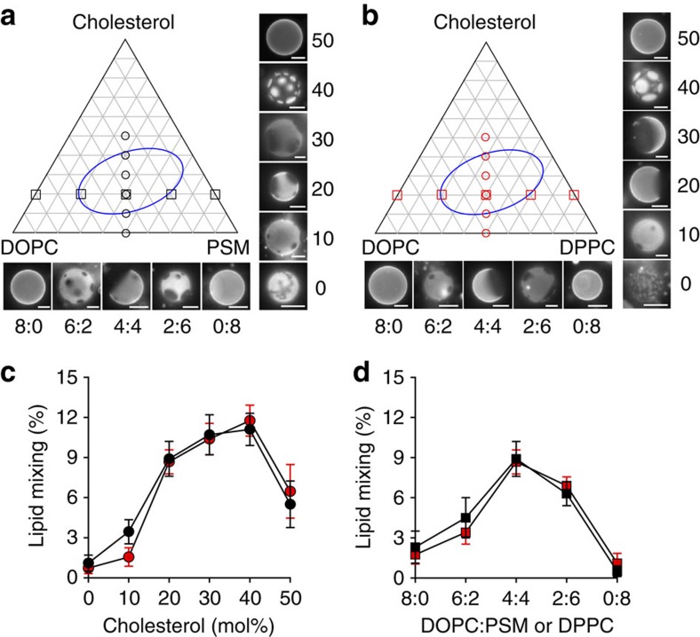
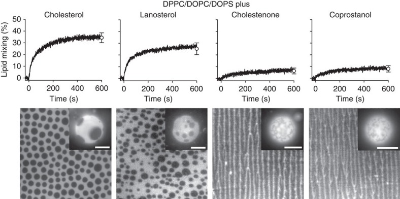
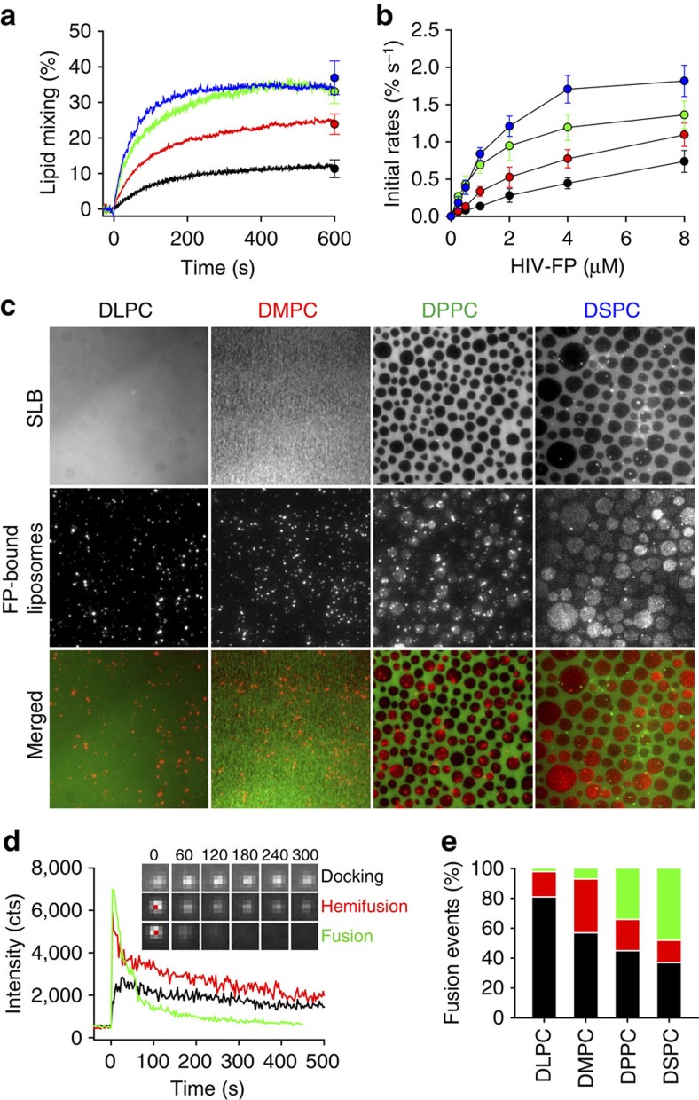
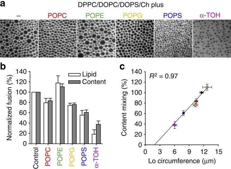
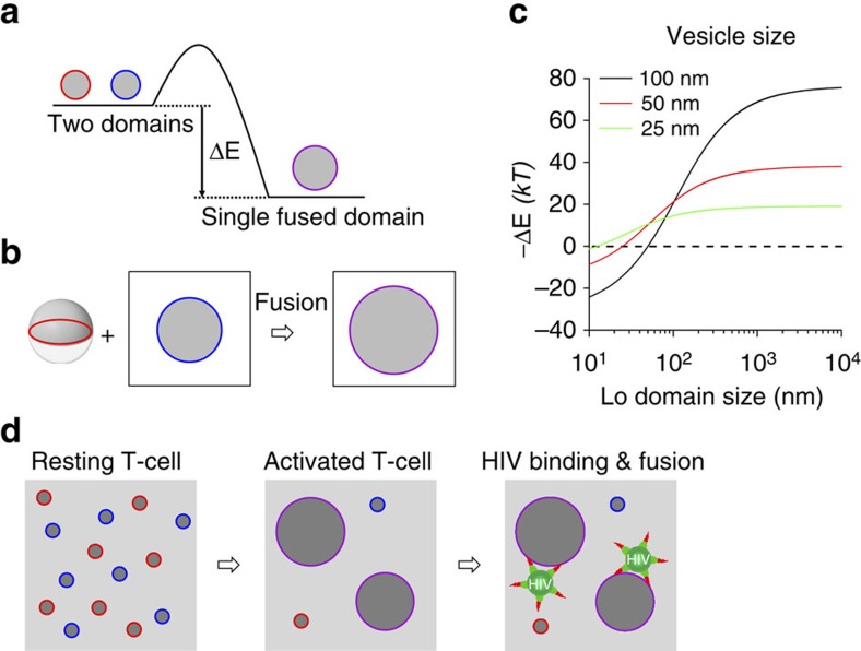
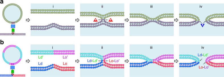
Lipids and proteins are organized in cellular membranes in clusters, often called 'lipid rafts'. Although raft-constituent ordered lipid domains are thought to be energetically unfavourable for membrane fusion, rafts have long been implicated in many biological fusion processes. For the case of HIV gp41-mediated membrane fusion, this apparent contradiction can be resolved by recognizing that the interfaces between ordered and disordered lipid domains are the predominant sites of fusion. Here we show that line tension at lipid domain boundaries contributes significant energy to drive gp41-fusion peptide-mediated fusion. This energy, which depends on the hydrophobic mismatch between ordered and disordered lipid domains, may contribute tens of kBT to fusion, that is, it is comparable to the energy required to form a lipid stalk intermediate. Line-active compounds such as vitamin E lower line tension in inhomogeneous membranes, thereby inhibit membrane fusion, and thus may be useful natural viral entry inhibitors.


Figures

Figure 1
Figure 1. Experimental design to study effects of lipid phase boundaries on HIV gp41-mediated membrane fusion.
Schematic representation of HIV gp41 interaction with Lo/Ld domain phase boundary. The fusion peptide of gp41 preferentially inserts and promotes membrane fusion at the interface between Lo and Ld phases. To address what molecular and physical properties are responsible for membrane fusion at these boundaries, we systematically modulated the interfaces by changing components of the Lo domains, modifying hydrophobic mismatch and introducing line-active molecules.
Figure 2
Figure 2. Comparison of the effect of Lo phase-promoting PSM and DPPC on lipid mixing mediated by HIV-FP.
Lipid mixing with 1 μM HIV-FP added to 50 μM LUVs composed of from left to right DOPC/DOPS (3:1), PSM/DOPC/DOPS/Ch (2:1:1:1), DPPC/DOPC/DOPS/Ch (2:1:1:1), PSM/DPPS/Ch (2:1:1) and DPPC/DPPS/Ch (2:1:1; top row). Fluorescence micrographs of GUVs with corresponding lipid compositions and labelled with 0.1 mol% Rh-PE (bottom row). Scale bars, 10 μm. Error bars are s.d. of three replicates.
Figure 3
Figure 3. Lipid phase-dependent membrane fusion mediated by HIV-FP.
Ternary lipid mixtures composed of (a) PSM/DOPC/Ch or (b) DPPC/DOPC/Ch with variable ratios were prepared as indicated in the triangular phase diagrams. Fluorescence micrographs of GUVs at constant 1:1 saturated:unsaturated lipid ratio with increasing cholesterol concentrations (0, 10, 20, 30, 40 and 50 mol% on perpendicular axes of the phase diagrams) and GUVs at constant 20 mol% cholesterol with variable DOPC/PSM (or DPPC) ratios (on horizontal axes of the phase diagrams). Scale bars, 10 μm. (c) Cholesterol- or (d) DOPC/PSM (or DPPC) ratio-dependent lipid mixing of 100 μM LUVs induced by 5 μM HIV-FP. LUVs are composed of the same lipid mixtures as used in a,b. PSM/DOPC/Ch and DPPC/DOPC/Ch data are shown with black and red symbols, respectively. Data are mean±s.d. from three experiments.
Figure 4
Figure 4. Effect of different sterols on Lo domain formation and membrane fusion.
Lipid mixing with 1 μM HIV-FP added to 50 μM LUVs composed of DPPC/DOPC/DOPS/sterol (2:1:1:1; top row). Sterols from left to right are cholesterol, lanosterol, cholestenone and coprostanol as indicated. Fluorescence micrographs of supported lipid monolayers and GUVs (insets) with corresponding lipid compositions and labelled with 0.1 mol% Rh-PE (bottom row). The scale of all lipid monolayers is 60 × 60 μm2 and scale bars in insets are 10 μm. Error bars are s.d. of three replicates.
Figure 5
Figure 5. Effect of hydrophobic mismatch on Lo domain formation and membrane fusion.
(a) Effect of saturated lipid component on lipid mixing. 1 μM HIV-FP was added to 50 μM LUVs composed of DSPC/DOPC/DOPS/Ch (2:1:1:1; blue), DPPC/DOPC/DOPS/Ch (2:1:1:1; green), DMPC/DOPC/DOPS/Ch (2:1:1:1; red) and DLPC/DOPC/DOPS/Ch (2:1:1:1; black). (b) Initial rates of lipid mixing as function of HIV-FP concentration. Same colour designations are used as in a. Data are mean±s.d. from three experiments. (c) Fusion between LUVs and SLB. LUVs and SLBs were composed of saturated lipid/DOPC/DOPS/Ch (2:1:1:1). The saturated lipids are DLPC, DMPC, DPPC or DSPC as indicated. LUVs were added to SLBs which were pre-incubated with 5 μM HIV-FP for 10 min. The images were acquired 20 min after vesicle addition. Fluorescence micrographs of SLBs labelled with 0.1 mol% Rh-PE (top row), TIRF micrographs of bound/fused LUVs labelled with 0.5 mol% DiD on SLB (middle row), and merged images (bottom row). The scale of all images is 64 × 64 μm2. (d) Representative single-LUV fusion events on SLBs including docking, hemifusion and full fusion. Time zero is defined as the first frame with a visible liposome. The insets show TIRF microscopy images of representative times (s) for each type of fusion event. The scale of all inset images is 2.5 × 2.5 μm2. (e) Relative frequencies of single-LUV docking (black), hemifusion (red) and full fusion (green) events.
Figure 6
Figure 6. Effect of linactants on Lo domain formation and membrane fusion.
(a) Fluorescence micrographs of supported lipid monolayers composed of DPPC/DOPC/DOPS/Ch (2:1:1:1) (control) with 20 mol% linactants (hybrid phospholipids and α-TOH) as indicated. The statistics of several observed parameters on the Lo domains as calculated by ImageJ are shown in Supplementary Fig. 3a. The scale of all images is 40 × 40 μm2. (b) Effect of linactants on membrane fusion mediated by HIV-FP. The extent of lipid (white bars) and content (grey bars) mixing was measured 10 min after addition of 1 μM HIV-FP to 50 μM LUVs composed of the same lipid mixtures as in a. (c) The dependence of content mixing on the circumference of Lo domains shows a linear relationship (correlation coefficient R2=0.97). The coloured data points correspond to the colours of the added linactants in a,b. The direct correlation of content (or lipid) mixing with the circumference of the domains indicates a critical role of the Lo/Ld interface line tension in membrane fusion. Data are mean±s.e.m. from three experiments.
Figure 7
Figure 7. Release of boundary energy by domain coalescence.
(a) Schematic diagram illustrating the change of boundary energy by domain coalescence. (b) A symmetric vesicle with two equally sized Lo- and Ld-phase hemispheres fuses with a Lo domain in a planar membrane. (c) Change of boundary energy for the reaction shown in b as a function of Lo domain size in the planar membrane for vesicles of different sizes. Domain fluctuations are not considered in this simple geometrical model (see text). (d) Schematic diagram illustrating biological implications of domain coalescence in T-cell activation on HIV binding and fusion at domain boundaries. The small domains shown in resting T-cells may actually be dynamic fluctuating clusters or nanodomains of receptors and lipids, but even then, the concept of lateral assembly of multiple clusters and domain growth during T-cell activation is still valid.
Figure 8
Figure 8. Extension of stalk-pore model of membrane fusion to heterogeneous lipid bilayers containing lipid rafts.
(a) Schematic diagrams illustrating different steps of membrane fusion in the standard stalk-pore model with homogeneous membranes: (i) close contact of two lipid bilayers with point-like protrusion, (ii) lipid stalk connecting two bilayers, (iii) hemifusion diaphragm and (iv) fusion pore. Lipids with negative spontaneous curvature (red triangles) stabilize the stalk and lipids with positive spontaneous curvature (blue inverted triangles) stabilize the fusion pore. (b) Stalk-pore model extended to fusion of lipid bilayers with coexisting Lo/Ld domains. The domain boundaries generate additional energy for membrane fusion by reduction of line tension energy. These boundaries induce local curvature and defects that facilitate fusion peptide insertion at different steps in the extended stalk-pore model (see text, for more detail).

References

    1. Doms R. W. & Moore J. P. HIV-1 membrane fusion: targets of opportunity. J. Cell Biol. 151, F9–F14 (2000). - PMC - PubMed
    1. Wilen C. B., Tilton J. C. & Doms R. W. HIV: cell binding and entry. Cold Spring Harb. Perspect. Med. 2, a006866 (2012). - PMC - PubMed
    1. Harrison S. C. Viral membrane fusion. Virology 479–480, 498–507 (2015). - PMC - PubMed
    1. Tamm L. K. & Han X. Viral fusion peptides: a tool set to disrupt and connect biological membranes. Biosci. Rep. 20, 501–518 (2000). - PubMed
    1. Tamm L. K., Lee J. & Liang B. Capturing glimpses of an elusive HIV gp41 prehairpin fusion intermediate. Structure 22, 1225–1226 (2014). - PMC - PubMed

Publication types

LinkOut - more resources