. 2016 Dec 1;80(11):827-835.
doi: 10.1016/j.biopsych.2016.03.1050. Epub 2016 Mar 10.

Genetic Disruption of Circadian Rhythms in the Suprachiasmatic Nucleus Causes Helplessness, Behavioral Despair, and Anxiety-like Behavior in Mice

Affiliations

Genetic Disruption of Circadian Rhythms in the Suprachiasmatic Nucleus Causes Helplessness, Behavioral Despair, and Anxiety-like Behavior in Mice

Dominic Landgraf et al. Biol Psychiatry. .

Abstract

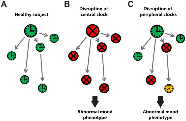
Background: Major depressive disorder is associated with disturbed circadian rhythms. To investigate the causal relationship between mood disorders and circadian clock disruption, previous studies in animal models have employed light/dark manipulations, global mutations of clock genes, or brain area lesions. However, light can impact mood by noncircadian mechanisms; clock genes have pleiotropic, clock-independent functions; and brain lesions not only disrupt cellular circadian rhythms but also destroy cells and eliminate important neuronal connections, including light reception pathways. Thus, a definitive causal role for functioning circadian clocks in mood regulation has not been established.

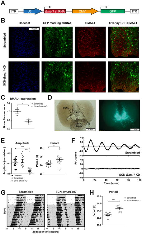
Methods: We stereotactically injected viral vectors encoding short hairpin RNA to knock down expression of the essential clock gene Bmal1 into the brain's master circadian pacemaker, the suprachiasmatic nucleus (SCN).

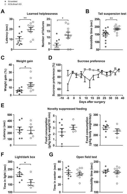
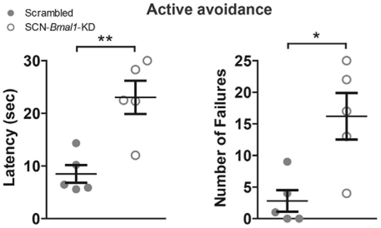
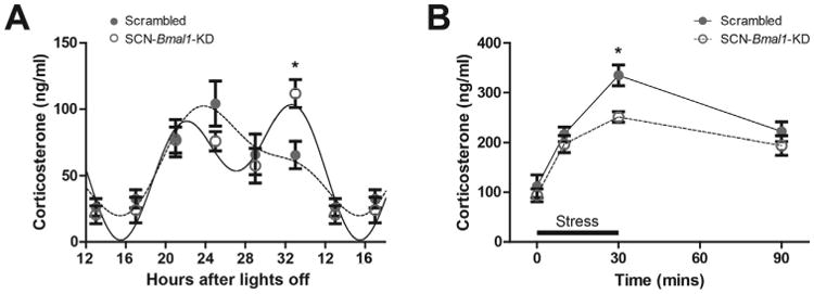
Results: In these SCN-specific Bmal1-knockdown (SCN-Bmal1-KD) mice, circadian rhythms were greatly attenuated in the SCN, while the mice were maintained in a standard light/dark cycle, SCN neurons remained intact, and neuronal connections were undisturbed, including photic inputs. In the learned helplessness paradigm, the SCN-Bmal1-KD mice were slower to escape, even before exposure to inescapable stress. They also spent more time immobile in the tail suspension test and less time in the lighted section of a light/dark box. The SCN-Bmal1-KD mice also showed greater weight gain, an abnormal circadian pattern of corticosterone, and an attenuated increase of corticosterone in response to stress.

Conclusions: Disrupting SCN circadian rhythms is sufficient to cause helplessness, behavioral despair, and anxiety-like behavior in mice, establishing SCN-Bmal1-KD mice as a new animal model of depression.

Keywords: Circadian clocks; Corticosterone; Depression; Learned helplessness; Mouse model; Suprachiasmatic nucleus.


Figures

Figure 1
Figure 1
Knockdown of Bmal1 expression abolishes circadian rhythms in suprachiasmatic nucleus (SCN). (A) Adeno-associated virus (AAV) expression constructs encoding green fluorescent protein (GFP) and short hairpin RNAs (shRNAs) targeting Bmal1. (B) SCN of mice were injected with AAVs carrying Bmal1-knockdown (Bmal1-KD) or scrambled shRNA sequences as well as a GFP reporter. Fluorescence images of representative fields show cell nuclei marked by Hoechst staining (blue), transduced cells marked by GFP (green), and BMAL1 protein levels marked by immunolabeling (red) from SCN-Bmal1-KD and control mice. The overlay shows that most Bmal1-shRNA transfected cells show reduced BMAL1 levels. (C) Immunofluorescence labeling reveals an ∼60% reduction of BMAL1 protein levels in the SCN of mice injected with Bmal1-shRNA relative to control mice. Data are shown as mean ± SEM; *p ≤ .05, t4 = 3.657 (Student t test); n = 3. (D) After AAV injections and subsequent behavioral experiments, coronal organotypic SCN explants of all mice were prepared (left) to confirm the correct location of injections based on GFP expression patterns (right) and to determine the efficiency of Bmal1 knockdown based on amplitude of mPer2Luciferase (PER2∷LUC) rhythms. (E) On average, knockdown of Bmal1 expression reduces PER2∷LUC rhythm amplitude by ∼80% and significantly lengthens the PER2∷LUC circadian rhythm period. SCN explants from mice that received scrambled sequence control injections show PER2∷LUC rhythms similar in amplitude to PER2∷LUC rhythms from untreated mice. Amplitude data are shown as mean ± SEM; F22,6 = 15.98 (one-way analysis of variance with Bonferroni posttest; ***p ≤ .001); n = 9–10. Period data are shown as mean ± SEM; t17 = 2.279 (Student t test); n = 9–10. (F) Representative PER2∷LUC rhythms of SCN explants injected with AAV particles encoding scrambled shRNA sequences (top) and shRNA targeting Bmal1 RNA (bottom). (G) Representative actograms showing wheel-running activity during entrainment in a 12:12 light/dark cycle and subsequent constant darkness. Gray shading represents times of darkness. (H) SCN-Bmal1-KD mice display longer circadian free-running periods of locomotor activity in constant darkness. Data are shown as mean ± SEM; **p ≤ .01, t11 = 4.104 (Student t test); scrambled, n = 8; SCN-Bmal1-KD, n = 5. CMV, cytomegalovirus; ITR, inverted terminal repeat.
Figure 2
Figure 2
Disruption of circadian rhythms in the suprachiasmatic nucleus (SCN) leads to increased helplessness, despair, weight gain, and anxiety-related behavior. (A) In the learned helplessness paradigm, Bmal1-knockdown (Bmal1-KD) in SCN increases escape latency times (left) and number of escape failures (right). Data are shown as mean 6 SEM; *p ≤ .05, **p ≤ .01; latency time, t17 = 2.997; escape failures, t17 = 2.801 (Student t test); n = 9–10. (B) In the tail suspension test, Bmal1-KD in SCN increases immobility time. Data are shown as mean ± SEM; **p ≤ .01; t25 = 3.067 (Student t test); n = 12–15. (C) During the 5 weeks after adeno-associated virus injection, SCN-Bmal1-KD mice gain significantly more weight than control mice. Data are shown as mean ± SEM; *p ≤ .05; t16 = 2.693 (Student t test); n = 8–10. (D) In the sucrose preference test, Bmal1-KD in SCN has no significant impact on preference for sweet water. Data are shown as mean 6 SEM; interaction, F10,120 = 1.189, p = .352; time, F1,120 = 1.355, p = .2093; genotype, F10,120 = .8537, p = .3737; post hoc test, not significant (two-way repeated measures analysis of variance with Bonferroni posttest); n = 9–10. (E) Suppression of SCN rhythms does not change the aversion to eating in a novel environment (expressed as latency to begin eating) or total food intake (expressed per 10 minutes during the test or per day). Data are shown as mean ± SEM; not significant; latency time, t11 = 0.1600; food intake in novel environment, t11 = 0.9643; daily food intake, t11 = 1.173 (Student t test); n = 5–8. (F) SCN-Bmal1-KD mice spend significantly less time in the light compartment of a light/dark box, which is conventionally interpreted as an increase in anxiety-related behavior. Data are shown as mean ± SEM; *p ≤ .05; t11 = 2.430 (Student t test); n = 5–8. (G) In the open field test, Bmal1-KD in SCN does not alter spatial preference or total activity. Data are shown as mean ± SEM; not significant; time in center, t15 = 0.5319; immobility time, t15 = 0.4397 (Student t test); n = 7–10.
Figure 3
Figure 3
Disruption of suprachiasmatic nucleus (SCN) circadian clock function leads to a state of helplessness as manifested by decreased active avoidance of foot shock. Even without previous exposure to inescapable stress, Bmal1-knockdown (Bmal1-KD) in SCN leads to higher escape latency times (left) and increased numbers of escape failures during testing in the learned helplessness shuttle boxes. Data are shown as mean ± SEM; *p ≤ .05, **p ≤ .01; latency time, t8 = 4.069; escape failures, t8 = 3.301 (Student t test); n = 5.
Figure 4
Figure 4
Suprachiasmatic nucleus–specific Bmal1-knockdown (SCN-Bmal1-KD) mice have altered stress hormone function. (A) Circadian patterns of corticosterone release in SCN-Bmal1-KD and control mice kept in constant darkness. In addition to the normal increase of corticosterone at the beginning of the subjective night (25 hours after “lights off”), SCN-Bmal1-KD mice have a second corticosterone peak at the end of subjective night (33 hours after “lights off”). Data are shown as mean ± SEM and Fourier-curve fits with two harmonics; interaction, F5,70 = 3.279, p = .0102; stress, F1,70 = 18.45, p ≤ .0001; genotype, F5,70 = 0.01066, p = .9192; post hoc test, *p ≤ .05 (two-way repeated measures analysis of variance with Bonferroni posttest); n = 8. (B) Corticosterone release of SCN-Bmal1-KD and control mice in response to acute restraint stress for 30 minutes. SCN-Bmal1-KD mice show an attenuated corticosterone increase. Data are shown as mean ± SEM; interaction, F3,39 = 1.23, p = .3101; stress, F1,39 = 38.45, p ≤ .0001; genotype, F3,39 = 6.500, p = .0242; post hoc test, *p ≤ .05 (two-way repeated measures analysis of variance with Bonferroni posttest); n = 7–8.
Figure 5
Figure 5
Alternative models of how disruption of the clock network could lead to abnormal mood phenotypes. (A) In healthy subjects, the clock network is intact and synchronized (green), including the suprachiasmatic nucleus master pacemaker (big clock) and various peripheral clocks, including brain clocks and clocks in other tissues such as the adrenal (small clocks). (B) If the suprachiasmatic nucleus master pacemaker is rendered non-rhythmic (red), all downstream peripheral clocks, including clocks important for mood regulation, are disturbed as well because their component cellular circadian oscillators are no longer synchronized by the suprachiasmatic nucleus. This leads to abnormal mood. (C) An abnormal mood phenotype may occur when only a subset of peripheral clocks is disturbed. This may involve loss of rhythms as a result of asynchronous component cellular oscillators (red) or loss of synchronization with other tissues, such as adrenal (yellow).

Comment in

  • Sleep and Mood: Chicken or Egg?
    Ptáǒek LJ, Fu YH, Krystal AD. Ptáǒek LJ, et al. Biol Psychiatry. 2016 Dec 1;80(11):810-811. doi: 10.1016/j.biopsych.2016.09.012. Biol Psychiatry. 2016. PMID: 27968724 No abstract available.

Similar articles

Cited by

References

    1. Welsh DK, Takahashi JS, Kay SA. Suprachiasmatic nucleus: Cell autonomy and network properties. Annu Rev Physiol. 2010;72:551–577. - PMC - PubMed
    1. Hattar S, Kumar M, Park A, Tong P, Tung J, Yau KW, et al. Central projections of melanopsin-expressing retinal ganglion cells in the mouse. J Comp Neurol. 2006;497:326–349. - PMC - PubMed
    1. Husse J, Leliavski A, Tsang AH, Oster H, Eichele G. The light-dark cycle controls peripheral rhythmicity in mice with a genetically ablated suprachiasmatic nucleus clock. FASEB J. 2014;28:4950–4960. - PubMed
    1. Kiessling S, Sollars PJ, Pickard GE. Light stimulates the mouse adrenal through a retinohypothalamic pathway independent of an effect on the clock in the suprachiasmatic nucleus. PloS One. 2014;9:e92959. - PMC - PubMed
    1. Oster H, Damerow S, Kiessling S, Jakubcakova V, Abraham D, Tian J, et al. The circadian rhythm of glucocorticoids is regulated by a gating mechanism residing in the adrenal cortical clock. Cell Metab. 2006;4:163–173. - PubMed

Publication types

Substances