. 2016 Oct;73(20):3917-33.
doi: 10.1007/s00018-016-2232-z. Epub 2016 Apr 25.

Akt1 promotes stimuli-induced endothelial-barrier protection through FoxO-mediated tight-junction protein turnover

Affiliations

Akt1 promotes stimuli-induced endothelial-barrier protection through FoxO-mediated tight-junction protein turnover

Fei Gao et al. Cell Mol Life Sci. 2016 Oct.

Abstract

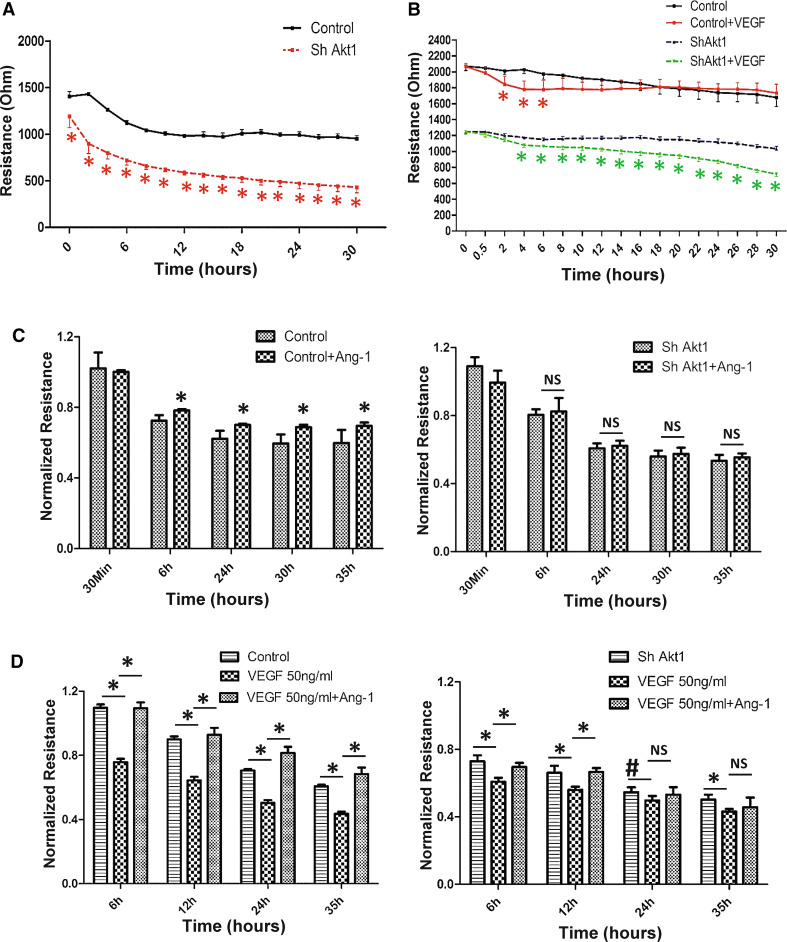
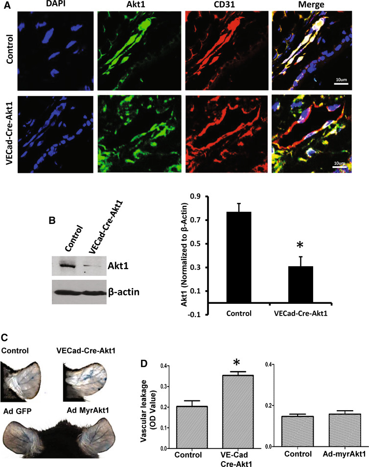
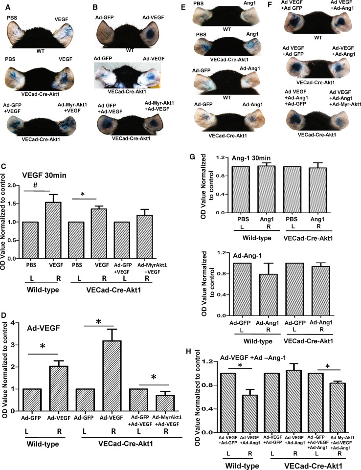
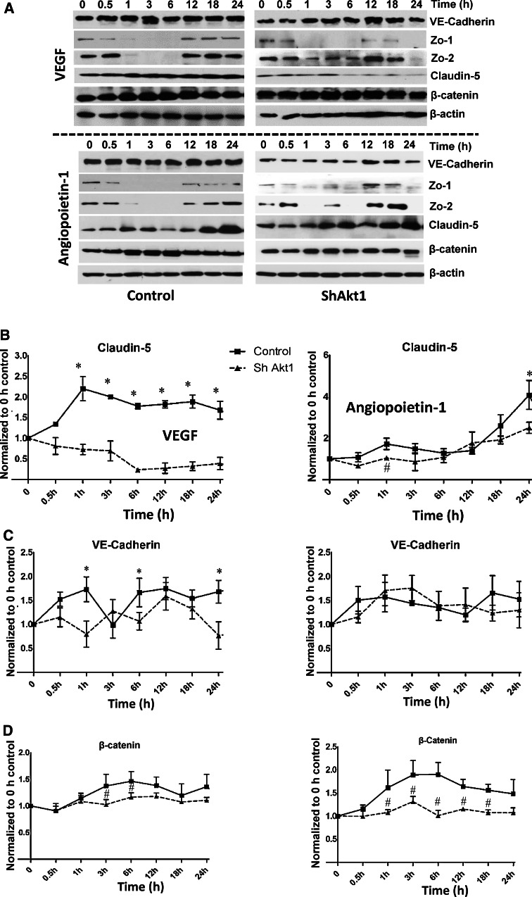
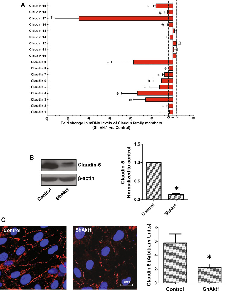
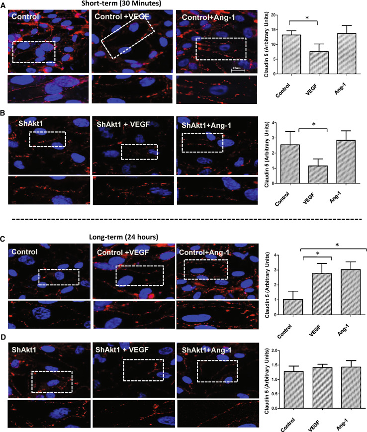
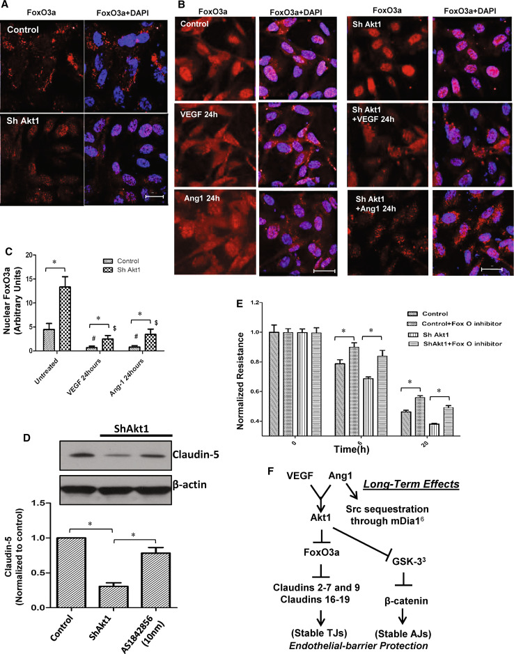
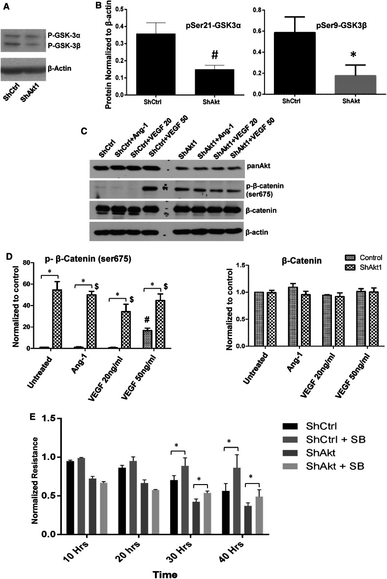
Vascular permeability regulated by the vascular endothelial growth factor (VEGF) through endothelial-barrier junctions is essential for inflammation. Mechanisms regulating vascular permeability remain elusive. Although 'Akt' and 'Src' have been implicated in the endothelial-barrier regulation, it is puzzling how both agents that protect and disrupt the endothelial-barrier activate these kinases to reciprocally regulate vascular permeability. To delineate the role of Akt1 in endothelial-barrier regulation, we created endothelial-specific, tamoxifen-inducible Akt1 knockout mice and stable ShRNA-mediated Akt1 knockdown in human microvascular endothelial cells. Akt1 loss leads to decreased basal and angiopoietin1-induced endothelial-barrier resistance, and enhanced VEGF-induced endothelial-barrier breakdown. Endothelial Akt1 deficiency resulted in enhanced VEGF-induced vascular leakage in mice ears, which was rescued upon re-expression with Adeno-myrAkt1. Furthermore, co-treatment with angiopoietin1 reversed VEGF-induced vascular leakage in an Akt1-dependent manner. Mechanistically, our study revealed that while VEGF-induced short-term vascular permeability is independent of Akt1, its recovery is reliant on Akt1 and FoxO-mediated claudin expression. Pharmacological inhibition of FoxO transcription factors rescued the defective endothelial barrier due to Akt1 deficiency. Here we provide novel insights on the endothelial-barrier protective role of VEGF in the long term and the importance of Akt1-FoxO signaling on tight-junction stabilization and prevention of vascular leakage through claudin expression.

Keywords: Akt; Angiopoietin-1; Claudin; VE-cadherin; VEGF; Vascular permeability.


Conflict of interest statement

Authors declare that there are no financial or conflicts of interests exist.

Figures

Fig. 1
Fig. 1
Akt1 deficiency compromises endothelial-barrier integrity and modulates VEGF and Ang-1-mediated endothelial-barrier function. a Real-time changes in the barrier resistance of ShControl and ShAkt1 HMEC monolayers as measured using ECIS equipment showing differences in endothelial-barrier resistance between ShControl and ShAkt1 HMEC monolayers at various time points after plating equal amount of cells in array wells (n = 3). b Graph showing quantification of a comparison between ShControl and ShAkt1 HMEC on the acute as well as chronic effects of optimal dose (20 ng/ml) of VEGF on barrier integrity (n = 3). c Bar graph showing quantification of a comparison between control and ShAkt1 knockdown HMEC on the acute as well as chronic effects of optimal dose of Ang-1 (50 ng/ml) on their barrier-junction integrity (n = 3). d Bar graph showing quantification of a comparison between control and ShAkt1 knockdown HMEC on the acute as well as chronic effects of supra-optimal dose of VEGF (50 ng/ml) alone and in combination with Ang-1 (50 ng/ml) on their barrier-junction integrity (n = 3). *P < 0.01
Fig. 2
Fig. 2
Endothelial specific Akt1 depletion lead to vascular leakage in vivo. a Confocal images showing specific knockdown of Akt1 in mouse endothelial cells (earlobe section), evidenced by the loss of Akt1 (green) in CD31 (red) positive blood vessels (n = 5). b Western blot images and optical densitometry analysis of aortic EC isolated from vehicle and tamoxifen treated VECad-Cre-Akt1 mice showing reduced Akt1 expression in tamoxifen-treated mice compared to untreated littermates (n = 3). c Representative images of the tamoxifen-treated WT and VECad-Cre-Akt1 (above), and Ad-GFP and Ad-myrAkt1 expressing WT mice (below) ears showing leakage of Evan’s blue dye. d Graph showing the quantification of leaked Evan’s blue dye (Miles assay) in tamoxifen-treated VECad-Cre-Akt1 knockout (left) and Ad-myrAkt1 expressing mice ears (right), compared to respective untreated control mice (n = 6). *P < 0.01 (scale bar 10 µM)
Fig. 3
Fig. 3
Akt1 augments VEGF-induced vascular permeability and Ang-1-induced vascular-barrier protection in vivo. a Representative images of PBS and 30 µl of 20 ng/ml recombinant VEGF administered WT mice (top), tamoxifen-treated VECad-Cre-Akt1 mice administered with PBS and VEGF (middle) and tamoxifen-treated VECad-Cre-Akt1 mice administered with Ad-GFP + VEGF and Ad-MyrAkt1 + VEGF ears for 30 min (short term) showing leakage of Evans blue dye (Miles assay). b Representative images of Ad-GFP and Ad-VEGF administered WT mice (top), tamoxifen-treated VECad-Cre-Akt1 mice administered with Ad-GFP and Ad-VEGF (middle) and tamoxifen-treated VECad-Cre-Akt1 mice ears expressing either Ad-GFP or Ad-myrAkt1 in the absence or presence of Ad-VEGF expression (long-term VEGF treatment) (bottom), showing leakage of Evans blue dye (Miles assay). c Histogram showing calorimetric quantification of the extravasated dye in the ears from PBS and 30 µl of 20 ng/ml recombinant VEGF administered WT mice (top), tamoxifen-treated VECad-Cre-Akt1 mice administered with PBS and VEGF (middle) and tamoxifen-treated VECad-Cre-Akt1 mice administered with Ad-GFP + VEGF and Ad-MyrAkt1 + VEGF after 30 min (n = 6). d Histogram showing calorimetric quantification of the extravasated dye in the ears from Ad-GFP and Ad-VEGF administered WT mice (top), tamoxifen-treated VECad-Cre-Akt1 mice administered with Ad-GFP and Ad-VEGF (middle) and tamoxifen-treated VECad-Cre-Akt1 mice ears expressing either Ad-GFP or Ad-myrAkt1 in the absence or presence of Ad-VEGF expression (long-term VEGF treatment) (n = 6). e Representative images of PBS and 30 µl of 50 ng/ml recombinant Ang1 administered WT mice and tamoxifen-treated VECad-Cre-Akt1 mice ears administered with PBS and 30 µl of 50 ng/ml recombinant Ang1 for 30 min (short term) (top two panels), and representative images of mice ears expressing Ad-GFP or Ad-Ang-1 (long term) in WT tamoxifen-treated VECad-Cre-Akt1 mice (bottom two panels) showing leakage of Evans blue dye (Miles assay). f Representative images of WT (top) and tamoxifen-treated VECad-Cre-Akt1 (middle) mice ears administered with combinations of either Ad-GFP/Ad-VEGF or Ad-VEGF/Ad-Ang-1, as well as VECad-Cre-Akt1 mice ears administered with a combination of either Ad-GFP/Ad-VEGF/Ad-Ang-1 or Ad-myrAkt1/Ad-VEGF/Ad-Ang-1 (bottom), showing leakage of Evans blue dye (Miles assay). g Histogram showing calorimetric quantification of the extravasated dye in the ears from WT and tamoxifen-treated VECad-Cre-Akt1 mice ears, with PBS or 30 µl PBS containing 50 ng/ml recombinant Ang-1 for 30 min, as well as WT and VECad-Cre-Akt1 mice ears, expressing either Ad-GFP or Ad-Ang-1 (n = 6). h Histogram showing calorimetric quantification of the extravasated dye in the ears from WT and tamoxifen-treated VECad-Cre-Akt1 mice administered with combinations of either Ad-GFP/Ad-VEGF or Ad-VEGF/Ad-Ang-1, as well as tamoxifen-treated VECad-Cre-Akt1 mice ears administered with a combination of either Ad-GFP/Ad-VEGF/Ad-Ang-1 or Ad-myrAkt1/Ad-VEGF/Ad-Ang-1 (n = 6). # P < 0.05, *P < 0.01
Fig. 4
Fig. 4
Akt1 deficiency affects real-time changes in the expression of proteins in endothelial-barrier AJs and TJs in response to VEGF and Ang-1 treatments. a Representative western blot images of control (left) and ShAkt1 (right) HMEC lysates treated with 20 ng/ml VEGF and real-time changes in the expression levels of AJ proteins VE-Cadherin and β-catenin as well as TJ proteins Zo-1, Zo-2 and claudin-5 were determined, and compared to 0 h time point (above; n = 3). Representative western blot images of control (left) and ShAkt1 (right) HMEC lysates treated with 50 ng/ml Ang-1 and real-time changes in the expression levels of AJ proteins VE-cadherin and β-catenin as well as TJ proteins Zo-1, Zo-2 and claudin-5 were determined, and compared to 0 h time point (below; n = 3). b Densitometry analysis of Western blot images of control and ShAkt1 HMEC lysates treated with 20 ng/ml VEGF (left) and 50 ng/ml Ang-1 (right) showing changes in claudin-5 expression compared to 0 h (n = 3). c Densitometry analysis of western blot images of control and ShAkt1 HMEC lysates treated with 20 ng/ml VEGF (left) and 50 ng/ml Ang-1 (right) showing changes in VE-cadherin expression compared to 0 h (n = 3). d Densitometry analysis of western blot images of control and ShAkt1 HMEC lysates treated with 20 ng/ml VEGF (left) and 50 ng/ml Ang-1 (right) showing changes in β-catenin expression compared to 0 h (n = 3). *P < 0.01, # P < 0.05
Fig. 5
Fig. 5
Long-term effect of Ang-1 and VEGF on endothelial-barrier protection is reliant on Akt1-mediated TJ stabilization. a Histogram showing fold-changes in mRNA levels of the claudin-family of TJ proteins with ShRNA-mediated Akt1 knockdown in HMEC, compared to control (n = 3). b Western blot showing reduced expression of endothelial claudin-5 in ShAkt1-HMEC compared to ShControl HMEC.  Right panel shows a bar graph with densitometry analysis of claudin-5 bands comparing ShControl and ShAkt1 HMEC lysates (n = 3). c Representative fluorescent images showing the expression of endothelial claudin-5 in control and ShAkt1 HMEC-barrier junctions as evidenced by fluorescence immunocytochemistry. Right panel shows bar graph comparing control and SkAkt1 HMEC monolayers for claudin-5 expression in their barrier junctions as detected by immunocytochemistry and quantified using NIH-Image J software (n = 5). *P < 0.01. (Scale bar 20 µM)
Fig. 6
Fig. 6
Long-term, not short-term changes in claudin-5 expression in HMEC is regulated by Akt1. a, b Representative images of claudin-5 staining of ShControl and ShAkt1 HMEC monolayers after 30 min (short-term) treatment with 20 ng/ml VEGF or 50 ng/ml Ang-1, compared to PBS control, respectively. Quantification of the claudin-5 expression levels in HMEC monolayers following VEGF and Ang-1 treatment as analyzed using NIH-Image J software is provided in the right panels (n = 10). c, d Representative images of claudin-5 staining of ShControl and ShAkt1 HMEC monolayers after 24 h (long-term) treatment with 20 ng/ml VEGF or 50 ng/ml Ang-1, compared to PBS control, respectively. Quantification of the claudin-5 expression levels in HMEC monolayers following VEGF and Ang-1 treatment as analyzed using NIH-Image J software is provided in the right panels (n = 10). *P < 0.01 (scale bar 20 µM)
Fig. 7
Fig. 7
Pharmacological inhibition of FoxO transcription factors restores endothelial-barrier function in ShAkt1 HMEC. a Confocal images of ShControl and ShAkt1 HMEC stained with FoxO3a antibodies and DAPI in the presence of FBS. b Confocal images of serum starved ShControl (left panel) and ShAkt1 (right panel) HMEC, pre-treated with PBS (top), VEGF (middle) or Ang-1 (bottom) for 24 h, and stained with FoxO3a antibodies and DAPI. c Bar graph showing quantification of the nuclear FoxO3a levels from the confocal images of ShControl and ShAkt1 HMEC, pre-treated with PBS, VEGF or Ang-1 for 24 h, and stained with FoxO3a antibodies and DAPI (n = 6). d Western blot analysis of lysates from ShControl and ShAkt1 HMEC prepared after 12 h treatment with 10 nM concentration of FoxO inhibitor AS1842856 (n = 3). Bar graph showing quantification of western blot analysis of ShControl and ShAkt1 HMEC prepared after 12 h treatment with 10 nM concentration of FoxO inhibitor AS1842856 is shown below (n = 4). e Bar graph showing the effect of FoxO inhibitor AS1842856 (10 nM) on endothelial-barrier resistance in ShControl and ShAkt1 HMEC as measured from the ECIS equipment (n = 4). f Working hypothesis on the role of Akt1 on acute and chronic vascular permeability. *P < 0.01 (scale bars 20 µM)
Fig. 8
Fig. 8
Pharmacological inhibition of GSK-3 in ShAkt1 HMEC partially restores the endothelial-barrier integrity in the long term. a, b Representative western blot images of ShControl and ShAkt1 HMEC lysates showing basal levels Ser9/21 phosphorylation of GSK-3α/β (n = 3). c Representative western blot images of ShControl and ShAkt1 HMEC lysates treated with PBS, 20 ng/ml VEGF, 50 ng/ml VEGF and 50 ng/ml Ang1 showing a comparison on phosphorylation and total expression of β-catenin, a GSK-3 substrate. d Densitometry analysis of Western blots of ShControl and ShAkt1 HMEC lysates treated with PBS, 20 ng/ml VEGF, 50 ng/ml VEGF and 50 ng/ml Ang1 showing a comparison on phosphorylation and total expression of β-catenin (n = 3). e Bar graph showing the effect of GSK-3 inhibitor SB415286 (20 µM) on endothelial-barrier resistance in ShControl and ShAkt1 HMEC as measured from the ECIS equipment (n = 4). *P < 0.01, # P < 0.05, $compared to untreated control

References

    1. Nagy JA, Dvorak AM, Dvorak HF. Vascular hyperpermeability, angiogenesis, and stroma generation. Cold Spring Harbor Perspect Med. 2012;2:a006544. doi: 10.1101/cshperspect.a006544. - DOI - PMC - PubMed
    1. Goddard LM, Iruela-Arispe ML. Cellular and molecular regulation of vascular permeability. Thromb Haemost. 2013;109:407–415. doi: 10.1160/TH12-09-0678. - DOI - PMC - PubMed
    1. Chen J, Somanath PR, Razorenova O, et al. Akt1 regulates pathological angiogenesis, vascular maturation and permeability in vivo. Nat Med. 2005;11:1188–1196. doi: 10.1038/nm1307. - DOI - PMC - PubMed
    1. Ha CH, Bennett AM, Jin ZG. A novel role of vascular endothelial cadherin in modulating c-Src activation and downstream signaling of vascular endothelial growth factor. J Biol Chem. 2008;283:7261–7270. doi: 10.1074/jbc.M702881200. - DOI - PMC - PubMed
    1. Daly C, Wong V, Burova E, et al. Angiopoietin-1 modulates endothelial cell function and gene expression via the transcription factor FKHR (FOXO1) Genes Dev. 2004;18:1060–1071. doi: 10.1101/gad.1189704. - DOI - PMC - PubMed

Publication types

MeSH terms

LinkOut - more resources