. 2016 May 5;165(4):813-26.
doi: 10.1016/j.cell.2016.04.010. Epub 2016 Apr 21.

Trimeric HIV-1-Env Structures Define Glycan Shields from Clades A, B, and G

Affiliations

Trimeric HIV-1-Env Structures Define Glycan Shields from Clades A, B, and G

Guillaume B E Stewart-Jones et al. Cell. .

Abstract

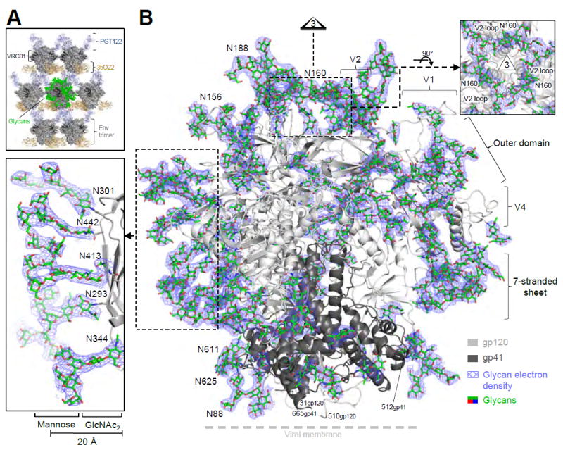
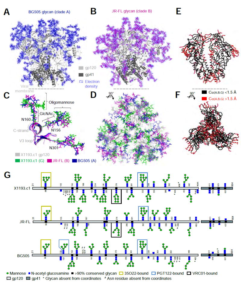
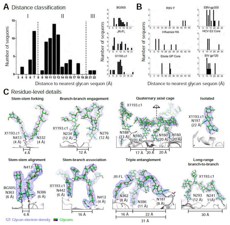
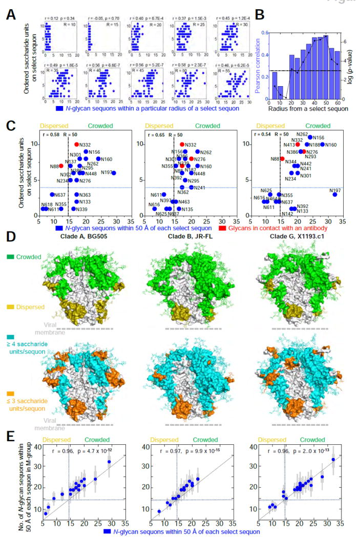
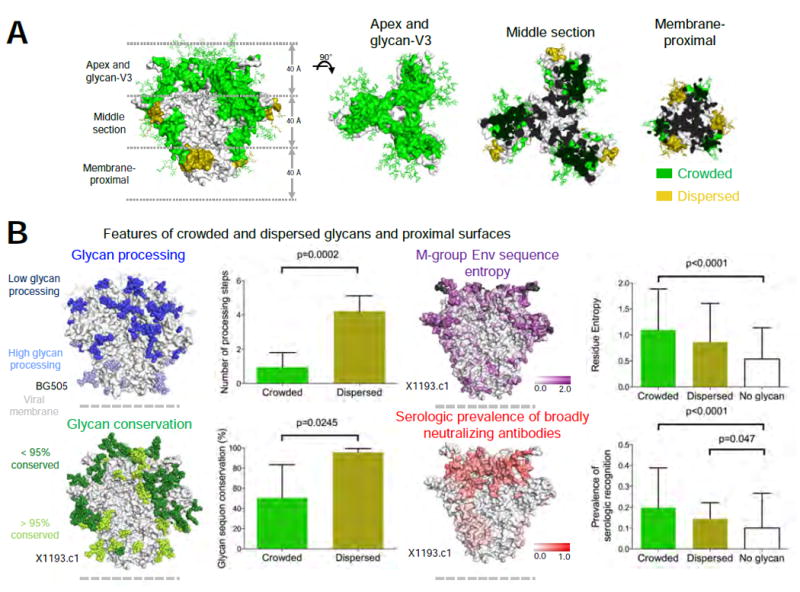
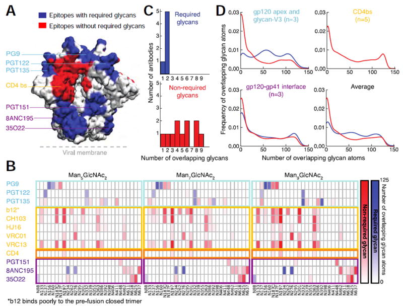
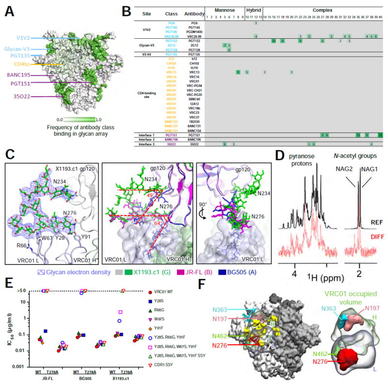
The HIV-1-envelope (Env) trimer is covered by a glycan shield of ∼90 N-linked oligosaccharides, which comprises roughly half its mass and is a key component of HIV evasion from humoral immunity. To understand how antibodies can overcome the barriers imposed by the glycan shield, we crystallized fully glycosylated Env trimers from clades A, B, and G, visualizing the shield at 3.4-3.7 Å resolution. These structures reveal the HIV-1-glycan shield to comprise a network of interlocking oligosaccharides, substantially ordered by glycan crowding, that encase the protein component of Env and enable HIV-1 to avoid most antibody-mediated neutralization. The revealed features delineate a taxonomy of N-linked glycan-glycan interactions. Crowded and dispersed glycans are differently ordered, conserved, processed, and recognized by antibody. The structures, along with glycan-array binding and molecular dynamics, reveal a diversity in oligosaccharide affinity and a requirement for accommodating glycans among known broadly neutralizing antibodies that target the glycan-shielded trimer.


Conflict of interest statement

COMPETING FINANCIAL INTERESTS

The authors declare no competing financial interests.

Figures

Figure 1
Figure 1. Crystal structure of a fully glycosylated HIV-1 Env trimer at 3.4 Å resolution reveals an ordered glycan shield
(A) Lattice comprising Fabs 35O22 (gold) and PGT122 (light blue) holds fully glycosylated HIV-1 Env trimers (surface representation in dark gray, with glycan highlighted in green on a representative trimer), with variable domain-only VRC01 in dark gray. (B) Crystal structure of a fully glycosylated SOSIP trimer from clade G strain X1193.c1 with gp120 (light gray) and gp41 (dark gray) in ribbon representation and glycans (green) in stick representation. 2Fo-Fc electron density (slate blue) is shown at 0.8 σ for glycans; altogether over half of the carbohydrate has been crystallographically resolved. Insets show select clusters of N-linked glycans, comprising N-acetylglucosamine residues protruding perpendicularly from the protein surface and supporting mannose branches, which form a glycan canopy ~20 Å from the protein surface. A view down the 3-fold is provided in Figure 2D. See also Figure S1 and Tables S3 and S4.
Figure 2
Figure 2. Fully glycosylated HIV-1 envelopes from clades A and B at 3.7 Å reveal conservation and diversity of protein and glycan shield
(A-B) HIV-1 trimers with protein in ribbon representation and glycan (BG505; blue, JR-FL; magenta) in sticks. 2Fo-Fc electron density (slate blue) is shown at 0.8 σ for glycans; assuming the average glycan is Man-7, ~50% of the glycan mass is ordered. (C-D) Superposition of Env trimers from clades A, B and G. Protein and glycan are colored as indicated, except that the clade G protein is shown in gray for clarity. (E-F) Env conserved core (black) with structural variation of Cα > 1.5 Å highlighted in red. (G) Residue-level schematic of crystallographically observed glycans. See also Figure S2 and Tables S3 and S4.
Figure 3
Figure 3. Taxonomy of glycan-glycan interactions that comprise the HIV-1 glycan shield
(A) Classification based on nearest interglycan sequon distance, with histograms showing glycans from each trimer (right) and from the composite of three trimers (left). (B) Nearest-neighbor analysis for various viral Env glycoproteins. (C) Glycan residue-level details for glycan-glycan interactions. Glycans are shown in stick representation, with 2Fo-Fc electron density (slate blue) at 0.8 σ. Nearest Nδ2 glycan sequon neighbor distance is shown in parentheses. See also Figure S3.
Figure 4
Figure 4. N-glycan crowding provides a mechanism for glycan order
(A) Number of crystallographically ordered saccharide units on a particular sequon relative to the density of neighboring N-linked glycans. Radii (R) range from 10-55 Å and are centered on Nδ2 of the Asn residue in the N-linked glycan sequon. There were a total of 69 occupied sequons in the three crystal structures. All correlation values were determined using a Pearson correlation (r). (B) Pearson correlation values (blue) and associated log(p-value) values (black) plotted for radii ranging from 5-60 Å. The dotted black line represents a p-value of 0.001 for reference. (C) The number of neighboring N-linked glycans observed in crystal structures for BG505 (clade A, left), JR-FL (clade B, middle) and x1193.c1 (clade G, right) at a radial cutoff of 50 Å. Blue circles denote sequon positions with at least one crystallographically observed saccharide unit. Red circles denote sequon positions with glycans in contact with an antibody. The total number of occupied sequons for BG505, JR-FL and X1193.c1 was 21, 23, and 25 respectively. The dotted lines represent the best partitioning of the x and y variables using Fishers Exact test (see methods for details). (D) Crowded and dispersed glycans mapped onto the surface of each Env trimer structure (top row). Crystallographically ordered glycans were also mapped onto the surface of each Env trimer structure (bottom row). N-linked glycans are displayed in stick representation. To enhance visualization, all neighboring residues within 10 Å were also colored accordingly. (E) For each glycan in the three crystallized strains, the corresponding glycan crowding was determined for each of 2994 M-group sequences by threading the BG505, JR-FL and X1193.c1 structures. Blue circles indicate the median glycan neighbors, thick grey bars indicate the interquartile range and thin grey bars indicate the 95% range around the median. We found significant correlation between the number of glycan neighbors in the three strains and the median neighbors for M-group sequences. See also Figure S4.
Figure 5
Figure 5. Biological characteristics of crowded and dispersed HIV-1 glycans and their associated surfaces
(A) Regions associated with crowded and dispersed glycans on the X1193.c1 trimer are shown from side (left panel) and top (three right panels) orientations, displayed as 40 Å sections through the trimer axis. (B) Features of crowded, dispersed and glycan-free surfaces. Correlations of crowded and dispersed glycans with glycan processing (upper left), glycan conservation (lower left), Env sequence entropy (upper right, insertion/deletion in hypervariable regions colored black) and serologic prevalence of broadly neutralizing antibodies (lower right). See also Figure S5E.
Figure 6
Figure 6. Molecular dynamics simulations reveal known broadly neutralizing antibodies that recognize the pre-fusion closed trimer need to accommodate N-linked glycan
(A) Epitopes for known broadly neutralizing antibodies displayed on the surface of the HIV-1 Env trimer. Epitopes are colored blue if N-linked glycans are known to be required for recognition and red if they are not required. (B) Glycan-antibody overlap analysis derived from MD simulations of Man-5, Man-7 and Man-9 models of the HIV-1 glycan shield. Overlap is colored blue for glycans known to be required for recognition by a particular antibody and red for glycans not known to be required for recognition. (C) Distribution graphs of required and non-required glycans. (D) Frequency of overlapping glycans versus number of overlapping glycan atoms. See also Figure S6, Table S6, and Movie S1.
Figure 7
Figure 7. Broadly neutralizing antibodies recognize oligosaccharides with a broad range of affinities
(A) Epitope-specific frequency of recognition of oligosaccharides by broadly neutralizing antibodies displayed on the surface of trimeric HIV-1 Env. (B) Relative fluorescence index (× 106) for broadly neutralizing antibody recognition of 40 oligosaccharides coupled to glass slides. (C) Ordered glycans N276 and N234 with 2Fo-Fc electron density at 0.8 σ in the X1193.c1 structure (left) bound to VRC01 light chain via van der Waal’s contacts and hydrogen bonds. Superimposition of BG505, JR-FL and X1193.c1 structures (middle) showing the relative difference in orientation of N276 and N234 glycans between the VRC01-bound X1193.c1 and JR-FL structures and the BG505 structure (which was not complexed to VRC01). (D) Detection of VCR01 binding to Man9GlcNAc2Asn by Saturation Transfer Difference (STD) NMR. Signals for sugar protons are labeled above the reference spectrum (REF) (glycan only, top). STD enhancements for both N-acetyl groups of the core GlcNAc residues and pyranose protons are displayed in the difference spectrum (DIFF) (VRC01:Man9GlcNAc2Asn complex, bottom). (E) Neutralization titers for VRC01 mutants at the N276 binding interface. (F) Molecular dynamics analysis of glycans overlapping the bound VRC01 volume, showing surrounding glycans and antibody on trimer (left) and isosurface representation of glycans overlapping VRC01 volume (right). See also Figure S7 and Table S6.

References

    1. Allan JS, Coligan JE, Barin F, McLane MF, Sodroski JG, Rosen CA, Haseltine WA, Lee TH, Essex M. Major glycoprotein antigens that induce antibodies in AIDS patients are encoded by HTLV-III. Science. 1985;228:1091–1094. - PubMed
    1. Correia BE, Bates JT, Loomis RJ, Baneyx G, Carrico C, Jardine JG, Rupert P, Correnti C, Kalyuzhniy O, Vittal V, et al. Proof of principle for epitope-focused vaccine design. Nature. 2014;507:201–206. - PMC - PubMed
    1. Crooks ET, Tong T, Chakrabarti B, Narayan K, Georgiev IS, Menis S, Huang X, Kulp D, Osawa K, Muranaka J, et al. Vaccine-Elicited Tier 2 HIV-1 Neutralizing Antibodies Bind to Quaternary Epitopes Involving Glycan-Deficient Patches Proximal to the CD4 Binding Site. PLoS Pathog. 2015;11:e1004932. - PMC - PubMed
    1. de Taeye SW, Ozorowski G, Torrents de la Pena A, Guttman M, Julien JP, van den Kerkhof TL, Burger JA, Pritchard LK, Pugach P, Yasmeen A, et al. Immunogenicity of Stabilized HIV-1 Envelope Trimers with Reduced Exposure of Non-neutralizing Epitopes. Cell. 2015;163:1702–1715. - PMC - PubMed
    1. Diskin R, Klein F, Horwitz JA, Halper-Stromberg A, Sather DN, Marcovecchio PM, Lee T, West AP, Jr, Gao H, Seaman MS, et al. Restricting HIV-1 pathways for escape using rationally designed anti-HIV-1 antibodies. J Exp Med. 2013;210:1235–1249. - PMC - PubMed

Publication types

Substances