. 2016 Jun 30;127(26):3398-409.
doi: 10.1182/blood-2015-10-674465. Epub 2016 Apr 25.

A thrombopoietin receptor antagonist is capable of depleting myelofibrosis hematopoietic stem and progenitor cells

Affiliations

A thrombopoietin receptor antagonist is capable of depleting myelofibrosis hematopoietic stem and progenitor cells

Xiaoli Wang et al. Blood. .

Erratum in

Abstract

Recently, interactions between thrombopoietin (TPO) and its receptor, the myeloproliferative leukemia (MPL) virus oncogene, have been shown to play a role in the development and progression of myeloproliferative neoplasms including myelofibrosis (MF). These observations have led to the development of strategies to disrupt the association of TPO with its receptor as a means of targeting MF hematopoietic stem cells (HSCs) and hematopoietic progenitor cells (HPCs). In this report, we show that although both splenic and peripheral blood MF CD34(+) cells expressed lower levels of MPL than normal CD34(+) cells, TPO promoted the proliferation of MF CD34(+) cells and HPCs in a dose-dependent fashion. Furthermore, the treatment of MF but not normal CD34(+) cells with a synthesized MPL antagonist, LCP4, decreased the number of CD34(+)Lin(-) cells and all classes of assayable HPCs (colony-forming unit-megakaryocyte [CFU-MK], CFU-granulocyte/macrophage, burst-forming unit-erythroid/CFU-erythroid, and CFU-granulocyte/erythroid/macrophage/MK) irrespective of their mutational status. In addition, LCP4 treatment resulted in the depletion of the number of MF HPCs that were JAK2V617F(+) Moreover, the degree of human cell chimerism and the proportion of malignant donor cells were significantly reduced in immunodeficient mice transplanted with MF CD34(+) cell grafts treated with LCP4. These effects of LCP4 on MF HSCs/HPCs were associated with inhibition of JAK-STAT activity, leading to the induction of apoptosis. These findings demonstrate that such specific anti-cytokine receptor antagonists represent a new class of drugs that are capable of targeting MF HSCs.


Figures

Figure 1
Figure 1
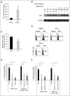
TPO concentrations in MF and normal plasmas, as well as MPL expression by MF and normal CD34+ cells. (A) TPO levels in MF and normal plasmas were measured using an enzyme-linked immunosorbent assay as described in supplemental Methods. The TPO concentration was significantly increased in MF plasma as compared with normal plasma. N plasma: n = 6; MF plasma: n = 13. (B) The expression of MPL by primary normal BM and MF CD34+ cells was determined by western blotting with a polyclonal Ab raised against the C-terminal amino acids of MPL. (C) Densitometric analysis of western blots as represented by (B) shows that the total level of MPL protein was reduced by 34% in MF CD34+ cells as compared with normal BM CD34+ cells. MF: n = 9; N BM: n = 6. (D-F) Expression of MPL on primary normal BM and MF CD34+ cells was evaluated by flow cytometry using a mAb, which specifically identifies the extracellular domain of MPL. (D) Representative flow cytometric plot showing MPL expression by normal BM (N BM14), MF splenic (SP19), and PB (PB195) CD34+ cells. (E) MPL expression was significantly reduced on MF splenic and PB CD34+ cells and CD34+Lin CD38 cells, as compared with normal BM counterparts. (F) Moreover, JAK2V617F+ and JAK2V617F MF CD34+ cells and CD34+LinCD38 cells showed a similar degree of reduction of MPL expression. *P < .05; **P < .01; ***P < .001. N BM: n = 8; MF splenic: n = 11; MF PB: n = 7; JAK2V617F+ MF: n = 9; and JAK2V617F MF: n = 9. N BM, normal BM; N plasma, normal plasma; SP, splenic.
Figure 2
Figure 2
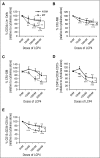
MF CD34+ cells are more sensitive to the effects of LCP4. (A-E) Splenic MF and normal BM CD34+ cells were cultured in the presence of cytokines (50 ng/mL SCF + 100 ng/mL TPO) alone or cytokines plus LCP4 at doses ranging from 10 nM to 500 nM for 1 week. After treatment, cells were analyzed by mAb staining and flow cytometry. A proportion of cells generated in these cultures were also assayed in semi-solid media to assess the effect of LCP4 on MF and normal HPCs. The percentage of the total number of CD34+Lin cells (A), CFU-MK (B), CFU-GM (C), as well as mature MKs (CD41a+CD34CD15) (D) and myeloid cells (CD15+CD34CD41a) (E), generated in the cultures exposed to cytokines plus LCP4 relative to those generated in the cultures exposed to cytokines alone is shown. *P < .05; **P < .01; ***P < .001; #P = .06; each dose of LCP4 vs 0 nM of LCP4 unless specified in the graphs. Splenic MF and N BM: n = 7 each. N BM, normal BM.
Figure 3
Figure 3
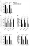
LCP4 treatment inhibits the proliferation and multilineage differentiation potential of MF splenic and PB CD34+ cells. MF splenic or PB CD34+ cells were treated with cytokines alone or cytokines plus LCP4 (100 nM, 500 nM) for 1 to 2 weeks. One week after the culture, the media were replaced with fresh media supplemented with or without LCP4 and cells were cultured for another week. Cells generated were phenotypically characterized and were assayed for HPCs. The percentage of the absolute number of CD34+Lin cells (A), all classes of assayable HPCs ([B] 1 week and [C] 2 weeks after the culture), as well as mature MKs (CD41a+CD34CD15) (D) and myeloid cells (CD15+CD34CD41a) (E), generated in the cultures of MF splenic or PB CD34+ cells exposed to cytokines plus LCP4 relative to that generated in the culture exposed to cytokines alone is shown. *P < .05; **P < .01; ***P < .001. n = 14 (8 splenic MF and 6 PB MF). WK, week.
Figure 4
Figure 4
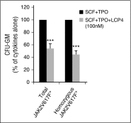
LCP4 treatment leads to the depletion of malignant HPCs. The absolute number of total JAK2V617F+ and homozygous JAK2V617F+ CFU-GM was calculated by multiplying the total number of CFU-GM by the fraction of JAK2V617F+ or homozygous JAK2V617F+ CFU-GM (Table 2), generated in cultures of JAK2V617F+ splenic or PB MF CD34+ cells treated with cytokines alone or cytokines plus LCP4 for 1 week. The percentage of the absolute number of total JAK2V617F+ and homozygous JAK2V617F+ CFU-GM generated in cultures of JAK2V617F+ splenic or PB MF CD34+ cells treated with cytokines plus LCP4, relative to that generated in cultures treated with cytokines alone is shown. n = 5. ***P < .001.
Figure 5
Figure 5
LCP4 treatment affects splenic MF stem cells but spares normal BM stem cells. (A-C) HCD45+ cell chimerisms in the BM (A), spleen (B), and PB (C) of individual mice receiving splenic MF or normal BM CD34+ cells treated with cytokines alone or cytokines plus LCP4 are shown. The percentages of hCD45+ in the BM, spleen, and PB of each individual mouse (♦) and the mean of the percentage of hCD45+ cells (horizontal bars) are shown. (D) The percentage of hCD45+ cell chimerism in the BM, spleen, and PB of mice receiving splenic MF or normal BM CD34+ cells treated with cytokines plus LCP4, relative to that detected in mice receiving cells treated with cytokines alone. **P < .01; ***P < .001. MF spleen: n = 6. SP10 and SP19: 3 mice transplanted; SP18, SP21, and SP22: 4 mice transplanted; and SP23: 2 mice transplanted due to the limited number of CD34+ cells available. N BM: n = 4, 2 mice transplanted for each sample due to the limited number of CD34+ cells available. Equal numbers of mice were transplanted with each MF spleen and normal BM sample with or without treatment with LCP4. N BM, normal BM; SP, splenic.
Figure 6
Figure 6
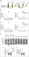
Mechanisms underlying the inhibitory effects of LCP4 treatment on MF CD34+ cells. (A-C) pSTAT3 and pSTAT5 levels in MF and normal BM CD34+ cells measured using phospho-flow cytometric analysis. (A) Representative flow cytometric plots showing pSTAT3 (left) and pSTAT5 (right) levels in MF splenic (SP14) and normal BM20 CD34+ cells. (B-C) Fold change in MFI of pSTAT3 (B) and pSTAT5 (C) for CD34+ cells from each MF spleen or normal BM, which was calculated using the equation: MFICytokines Alone / MFINo Cytokines or MFICytokines + LCP4 / MFINo Cytokines. Cytokines alone vs cytokines + LCP4: pSTAT3: MF and N BM: P for both > .05; pSTAT5: MF: P < .05; N BM: P > .05. (D) pMPL, pJAK2, pSTAT3, and pSTAT5 levels measured using western blotting in splenic MF CD34+ cells from 1 JAK2V617F and CALR mutation (SP14), 1 JAK2V617F but CALR mutation+ (SP15), and 2 JAK2V617F+ (SP9 and SP22) patients following treatment with cytokines alone or cytokines + LCP4. As indicated by arrows, LCP4 treatment resulted in the inhibition of pMPL, and/or pJAK2, pSTAT3/5 levels to varying degrees in both JAK2V617F+ and CALR mutation+ MF CD34+ cells, whereas only limited inhibition of TPO/MPL and JAK-STAT activity was observed with normal BM CD34+ cells. (E-F) Both the percentage (E) and absolute number (F) of CD34+ cells that were Annexin V+ and PI were greater in cultures of splenic MF CD34+ cells treated with cytokines plus LCP4 as compared with cells treated with cytokines alone. *P < .05. Splenic MF: n = 8; N BM: n = 6. N BM, normal BM.

References

    1. Klampfl T, Gisslinger H, Harutyunyan AS, et al. Somatic mutations of calreticulin in myeloproliferative neoplasms. N Engl J Med. 2013;369(25):2379–2390. - PubMed
    1. Nangalia J, Massie CE, Baxter EJ, et al. Somatic CALR mutations in myeloproliferative neoplasms with nonmutated JAK2. N Engl J Med. 2013;369(25):2391–2405. - PMC - PubMed
    1. Tefferi A, Lasho TL, Finke CM, et al. CALR vs JAK2 vs MPL-mutated or triple-negative myelofibrosis: clinical, cytogenetic and molecular comparisons. Leukemia. 2014;28(7):1472–1477. - PubMed
    1. Tefferi A. Myelofibrosis with myeloid metaplasia. N Engl J Med. 2000;342(17):1255–1265. - PubMed
    1. Yoshihara H, Arai F, Hosokawa K, et al. Thrombopoietin/MPL signaling regulates hematopoietic stem cell quiescence and interaction with the osteoblastic niche. Cell Stem Cell. 2007;1(6):685–697. - PubMed

Publication types

MeSH terms