. 2016 May 10;113(19):5376-81.
doi: 10.1073/pnas.1603335113. Epub 2016 Apr 25.

Targeting lymphoma with precision using semisynthetic anti-idiotype peptibodies

Affiliations

Targeting lymphoma with precision using semisynthetic anti-idiotype peptibodies

James Torchia et al. Proc Natl Acad Sci U S A. .

Abstract

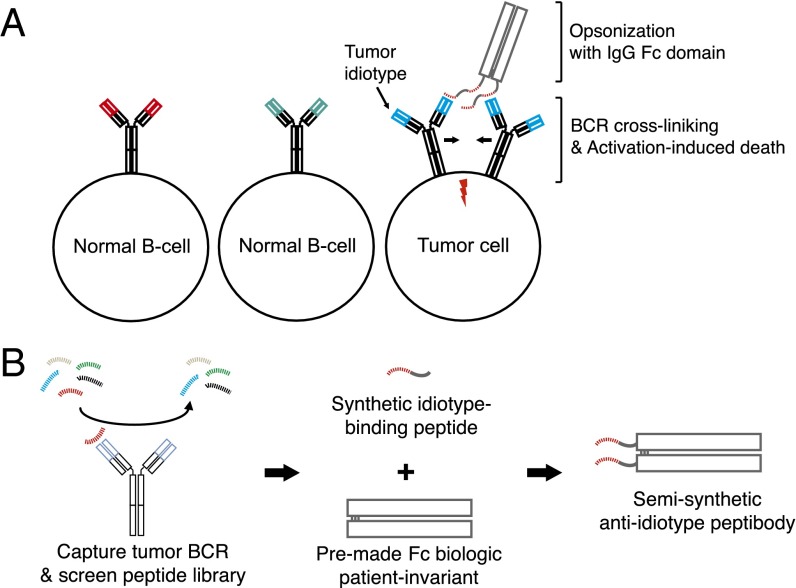
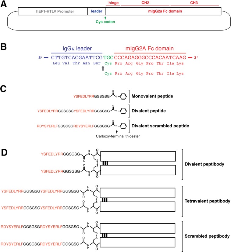
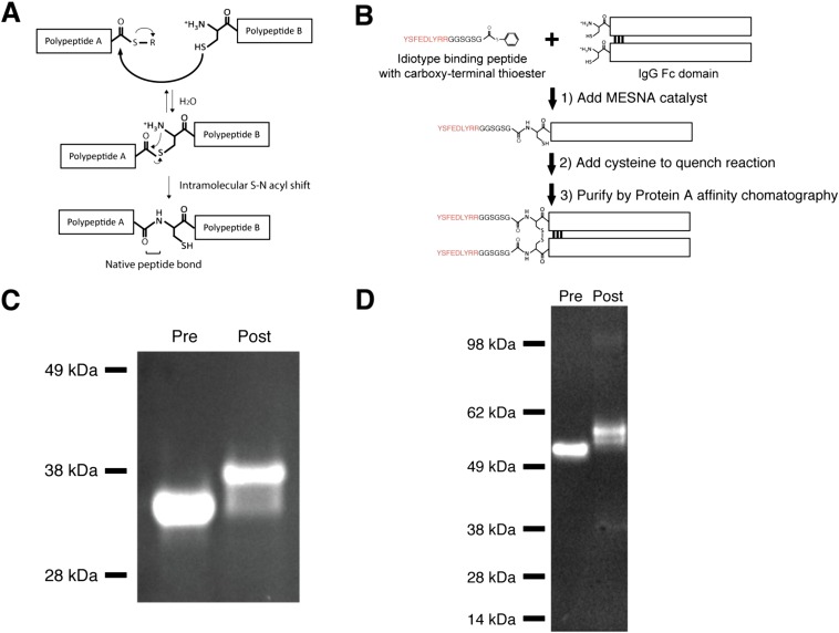
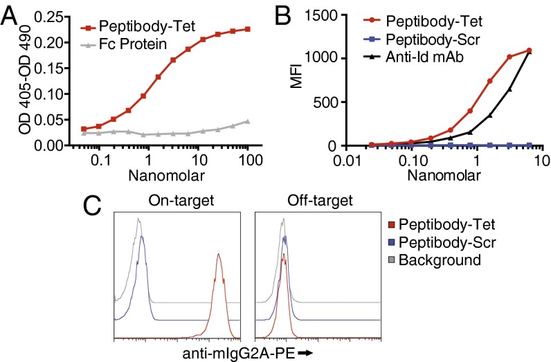
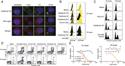
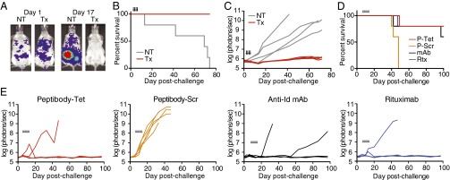
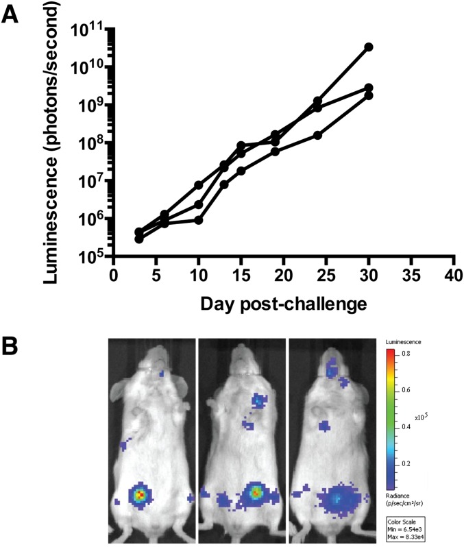
B-cell lymphomas express a functionally active and truly tumor-specific cell-surface product, the variable region of the B-cell receptor (BCR), otherwise known as idiotype. The tumor idiotype differs, however, from patient to patient, making it a technical challenge to exploit for therapy. We have developed a method of targeting idiotype by using a semisynthetic personalized therapeutic that is more practical to produce on a patient-by-patient basis than monoclonal antibodies. In this method, a small peptide with affinity for a tumor idiotype is identified by screening a library, chemically synthesized, and then affixed to the amino terminus of a premade IgG Fc protein. We demonstrate that the resultant semisynthetic anti-idiotype peptibodies kill tumor cells in vitro with specificity, trigger tumor cell phagocytosis by macrophages, and efficiently clear human lymphoma in a murine xenograft model. This method could be used to target tumor with true precision on a personalized basis.

Keywords: idiotype; lymphoma; peptibody; precision medicine.


Conflict of interest statement

The authors declare no conflict of interest.

Figures

Fig. 1.
Fig. 1.
Concept overview. (A) Tumor idiotype is the variable region of the BCR on the surface of lymphoma cells. It is unique to the tumor clone and distinct from the idiotype on other B-cell clones. Peptibodies cross-link idiotype, triggering BCR signaling, resulting in activation-induced cell death, and opsonize tumor cells with an IgG Fc domain, promoting tumor clearance mediated by innate immune effector cells. (B) Schematic of semisynthetic anti-idiotype peptibody production. A random peptide library is screened to identify peptide sequences with idiotype-specific binding, which are synthesized and affixed to the amino terminus of a premade recombinant IgG Fc domain.
Fig. S1.
Fig. S1.
Construct design. (A and B) Recombinant IgG Fc protein with an amino-terminal cysteine. (A) A human IgG kappa leader sequence was placed 5′ to nucleotide sequence encoding the mouse IgG2a hinge region, CH2 domain, and CH3 domain. A cysteine codon was inserted immediately 3′ to the leader sequence and 5′ to the mIgG2a sequence. (B) HEK 293 cells were stably transformed with this vector. Translation begins at a methionine codon on the 5′ end of the IgG kappa leader sequence and continues through a stop codon on the 3′ end of the mIgG2a sequence. The leader sequence is cleaved (arrow) by endogenous cellular endoproteases to reveal an amino-terminal cysteine on the secreted protein product. (C and D) Semisynthesis of peptibodies used in subsequent studies. (C) Synthetic peptides containing one or two idiotype binding sequences for the SUPB8 human lymphoma, or scrambled binding sequences, were produced by a commercial vendor with a carboxyl-terminal thioester moiety included during synthesis. These peptide thioesters were covalently linked to the amino terminus of a recombinant mouse IgG2a Fc domain by native chemical ligation. (D) The resultant peptibodies displayed either two (divalent peptibody) or four (tetravalent peptibody) idiotype-binding sequences, or four scrambled idiotype-binding sequences (scrambled peptibody).
Fig. S2.
Fig. S2.
Semisynthesis of peptibodies by NCL. (A) NCL reaction mechanism. The reaction proceeds in two steps: a reversible substitution of the carboxyl-terminal thioester moiety on one polypeptide for the sulfhydryl side chains of cysteine residues on the other polypeptide, followed by an irreversible intramolecular acyl shift that occurs selectively at the amino-terminal cysteine to yield a peptide bond. (B) Schematic of peptibody semisynthesis by NCL. A peptide with an idiotype-binding sequence and a carboxyterminal thioester moiety incorporated during synthesis are mixed in the presence of MESNA, which acts as both a reducing agent and catalyst. This results in ligation of the peptide to the amino terminus of dissociated Fc domain polypeptide chains. Free cysteine is then added to quench the reaction, and ligated Fc domain chains are purified from the reaction mixture by Protein A affinity chromatography. Once eluted into an oxidizing environment, Fc domain dimers reform. (C) Reducing SDS/PAGE gel of NCys-mIgG2a Fc protein alone (Pre) and semisynthetic peptibody (Post). (D) Nonreducing SDS/PAGE gel of NCys-mIgG2a Fc protein alone (Pre) and semisynthetic peptibody (Post).
Fig. 2.
Fig. 2.
Targeted idiotype and tumor binding. (A) Purified Ig from the on-target SUPB8 human lymphoma cells was immobilized on ELISA plates and exposed to varying concentrations of peptibody (Peptibody-Tet) or recombinant IgG2a Fc protein with no peptide attached. Binding was detected with an anti-mIgG2a-HRP antibody. The results are representative of at least four experiments. (B) On-target SUPB8 cells were exposed to varying concentrations of peptibody (Peptibody-Tet), peptibody with a scrambled idiotype peptide ligand sequence (Peptibody-Scr), or an anti-idiotype monoclonal antibody (Anti-Id mAb). Binding was detected with fluorophore-labeled anti-mIgG2a. Mean fluorescence intensity is plotted. The results are representative of at least four experiments. (C) On-target SUPB8 cells and off-target RAMOS cells with isotype-matched surface Ig were exposed to a saturating concentration of peptibody (Peptibody-Tet) or peptibody with a scrambled idiotype peptide ligand sequence (Peptibody-Scr). Binding was detected with fluorophore-labeled anti-mIgG2a. Results are representative of at least four experiments.
Fig. 3.
Fig. 3.
Anti-idiotype peptibodies cluster BCR, directly trigger BCR signaling and caspase-3 cleavage, and directly trigger tumor cell apoptosis and death. (A) On-target SUPB8 cells were treated with a saturating 100-nM concentration of peptibody tetramer (Peptibody-Tet) for 0 h (20 min on ice), 1 h, or 12 h at 37 °C. Cells were fixed with paraformaldehyde, permeabilized with cold methanol, and stained with anti-mIgG2a-FITC to detect bound peptibody (green) and anti-hIgM-APC to detect BCR (red). Cells were imaged with confocal microscopy, and representative images are shown. (B) Cells were incubated with a saturating 100-nM concentration of peptibody tetramer (Peptibody-Tet), peptibody dimer (Peptibody-Di), scrambled peptibody (Peptibody-Scr), anti-idiotype mAb (Anti-Id mAb), or anti-IgM F(ab’)2 for 5 min at 37 °C. Cells were fixed with paraformaldehyde, permeabilized with cold methanol, and stained with fluorophore-labeled anti-phospho Syk or anti-phospho ERK-1/-2. (C) Cells were incubated with mIgG2a Fc protein, peptibody, or anti-IgM Fab2’ for 24 h at 37 °C. Cells were fixed with paraformaldehyde, permeabilized with cold methanol, and stained with fluorophore-labeled anti-cleaved caspase-3. (D) Cells were incubated with 100 nM of peptibody tetramer, peptibody dimer, scrambled peptibody, anti-idiotype monoclonal antibody, or an anti-hIgM F(ab′)2 fragment for 24 h. Cell viability was assessed by 7-AAD exclusion, and surface phosphatidylserine expression was assessed by staining with FITC-labeled Annexin V. (E) Cells were incubated with varying concentrations of peptibody, mIgG2a Fc protein, anti-hIgM mAb, or anti-hIgG mAb for 3 d. Viability was assessed with the resazurin-based PrestoBlue dye. Fluorescence values were normalized to readings from untreated cells.
Fig. 4.
Fig. 4.
Anti-idiotype peptibodies promote innate immune effector-mediated clearance in vitro. (A) RFP+ mouse macrophages were incubated with GFP+ SUPB8 cells or GFP+ Raji cells in the presence of a saturating 100-nM concentration of peptide ligand, peptibody, mIgG2a Fc protein, anti-idiotype mAb, or rituximab. Representative FACS plots demonstrating RFP/GFP double-positive macrophages that have phagocytosed-labeled tumor cells. (B) Phagocytosis normalized to average % GFP+ macrophages upon treatment with anti-CD47 for each cell line (% maximal response). (C) Phagocytosis of SUPB8 cells in response to varying concentrations of peptibody. Normalized to mean percentage of GFP+ macrophages at saturating dose.
Fig. S3.
Fig. S3.
Idiotype binding peptide stability and peptibody pharmacokinetic profile. (A) Biotinylated idiotype binding peptide (YSFEDLYRRGGK-[biotin]) was added to mouse serum and incubated at 37 °C. Binding to purified idiotype at each timepoint was assessed by ELISA. (B and C) A CB-17 SCID mouse was injected i.p. with 50 μg of peptibody. Serum was obtained from tail bleeds at various timepoints. Peptibody concentration was determined at each time point by ELISA assessing mIgG2a concentration (B) and binding to purified idiotype (C).
Fig. 5.
Fig. 5.
Clearance of disseminated human lymphoma xenografts. (A–C) CB-17 SCID mice were challenged at day 0 with 1 × 10⁶ SUPB8-Luc+GFP+ cells i.v. and, beginning on day 2, were treated once daily with i.p. injections of 65 μg of peptibody for 4 d. Tumor burden was quantified by near-infrared imaging (Fig. S4). (A) Representative images of untreated (NT) and treated (Tx) mice at two timepoints. (B) Survival comparison. (C) Tumor burden quantified by near-infrared imaging. (D and E) CB-17 SCID mice were challenged at day 0 with 1 × 10⁶ SUPB8-Luc+GFP+ cells i.v. and, beginning on day 6, were treated once daily with i.p. injections of 50 μg of peptibody, scrambled peptibody, anti-idiotype mAb, or rituximab for 8 d. (D) Survival comparison. (E) Tumor burden quantified by near-infrared imaging.
Fig. S4.
Fig. S4.
SUPB8-Luc+GFP+ growth in SCID mice. (A) CB-17 SCID mice were challenged on day 0 with 1 × 10⁶ SUPB8-Luc+GFP+ cells i.v. (tail vein injection). Tumor burden was quantified by near-infrared imaging. (B) Images of mice on day 6 following challenge.

Similar articles

Cited by

References

    1. Shankland KR, Armitage JO, Hancock BW. Non-Hodgkin lymphoma. Lancet. 2012;380(9844):848–857. - PubMed
    1. Ng AK, LaCasce A, Travis LB. Long-term complications of lymphoma and its treatment. J Clin Oncol. 2011;29(14):1885–1892. - PubMed
    1. Morrison VA. Immunosuppression associated with novel chemotherapy agents and monoclonal antibodies. Clin Infect Dis. 2014;59(Suppl 5):S360–S364. - PubMed
    1. Zitvogel L, Apetoh L, Ghiringhelli F, Kroemer G. Immunological aspects of cancer chemotherapy. Nat Rev Immunol. 2008;8(1):59–73. - PubMed
    1. Shokat KM, Goodnow CC. Antigen-induced B-cell death and elimination during germinal-centre immune responses. Nature. 1995;375(6529):334–338. - PubMed

Publication types

MeSH terms