. 2016 May 10;113(19):5251-6.
doi: 10.1073/pnas.1525034113. Epub 2016 Apr 25.

Divergent non-heme iron enzymes in the nogalamycin biosynthetic pathway

Affiliations

Divergent non-heme iron enzymes in the nogalamycin biosynthetic pathway

Vilja Siitonen et al. Proc Natl Acad Sci U S A. .

Abstract

Nogalamycin, an aromatic polyketide displaying high cytotoxicity, has a unique structure, with one of the carbohydrate units covalently attached to the aglycone via an additional carbon-carbon bond. The underlying chemistry, which implies a particularly challenging reaction requiring activation of an aliphatic carbon atom, has remained enigmatic. Here, we show that the unusual C5''-C2 carbocyclization is catalyzed by the non-heme iron α-ketoglutarate (α-KG)-dependent SnoK in the biosynthesis of the anthracycline nogalamycin. The data are consistent with a mechanistic proposal whereby the Fe(IV) = O center abstracts the H5'' atom from the amino sugar of the substrate, with subsequent attack of the aromatic C2 carbon on the radical center. We further show that, in the same metabolic pathway, the homologous SnoN (38% sequence identity) catalyzes an epimerization step at the adjacent C4'' carbon, most likely via a radical mechanism involving the Fe(IV) = O center. SnoK and SnoN have surprisingly similar active site architectures considering the markedly different chemistries catalyzed by the enzymes. Structural studies reveal that the differences are achieved by minor changes in the alignment of the substrates in front of the reactive ferryl-oxo species. Our findings significantly expand the repertoire of reactions reported for this important protein family and provide an illustrative example of enzyme evolution.

Keywords: Streptomyces; crystal structure; iron-dependent oxygenase; natural product biosynthesis; α-ketoglutarate–dependent oxygenase.


Conflict of interest statement

The authors declare no conflict of interest.

Figures

Fig. 1.
Fig. 1.
The nogalamycin gene cluster and the biosynthetic steps for anthracyclines investigated in this study. The end product of the pathway nogalamycin (1) is produced by WT Streptomyces nogalater whereas pathway engineering led to the isolation of compounds 27 from the heterologous host S. albus, and compounds 8 and 9 were generated using enzymatic synthesis. The core of the gene cluster (white genes) has been cloned into cosmid pSnogaori for production of 2 whereas genes near the boundaries of the cluster are marked in black. Genes encoding SnoN and SnoK are shown in purple and gray, respectively.
Fig. S1.
Fig. S1.
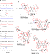
In vivo characterization of snoN and snoK. HPLC traces and extracted ion chromatogram have been recorded from culture extracts of (trace a) S. albus/pSnogaori+N, (trace b) S. albus/pSnogaori, and (trace c) S. albus/pSnoΔK. (A) HPLC-UV/Vis monitored at 470 nm. The extracted ion chromatogram by liquid chromatography-electrospray ionization-high resolution mass spectrometry (LC-ESI-HRMS) for (B) compounds 2 (m/z observed, 742.2727 [M-H]; calculated, 742.2716) and 3 (m/z observed, 742.2680 [M-H]; calculated, 742.2716), (C) compounds 4 (m/z observed, 698.2428 [M-H]; calculated, 698.2454) and 7 (m/z observed, 698.2435 [M-H]; calculated, 698.2454), (D) compound 5 (m/z observed, 744.2910 [M-H]; calculated, 744.2873), and (E) compound 6 (m/z observed, 700.2641 [M-H]; calculated, 700.2611). (F) Key HMBC signals (red arrows) and coupling constants (blue dashed line) for 4, 5, 6, and 7.
Fig. S2.
Fig. S2.
In vitro characterization of SnoK and SnoN. (A, Upper) The spectral properties of 5 (black) and 2 (red). (Lower) HPLC traces of the SnoK reactions at 470 nm from (trace a) a negative control reaction with 5, (trace b) SnoK reaction with 5 producing 2, and (trace c) an authentic standard for 2. (B) The oxygen dependency of SnoK. The reaction does not proceed if air is depleted (black), but 5 is converted into 2 after reintroduction of air to the reaction (red). (C) The Fe(II) dependency of SnoK, with EDTA (black) and without EDTA (red). (D) Titration of the SnoK (red) and SnoN (gray) reactions with varying concentrations of α-KG with the substrate 5 concentration kept at 50 µM. (E, Upper) The spectral properties of 7 (black) and 8 (red). (Lower) HPLC traces of the SnoN reactions at 470 nm from (trace a) a negative control reaction with 7, (trace b) SnoN reaction with 7 producing 8, and (trace c) purified standard 8 characterized by NMR in this study. (F) Key HMBC signals and HRMS data for 8. (G, Upper) The spectral properties of 5 (black) and 9 (red). (Lower) HPLC traces of the SnoN reactions at 470 nm from (trace a) a negative control reaction with 5, (trace b) SnoN reaction with 5 producing 9, and (trace c) an authentic standard for 9 obtained from previous studies (9). (H) The oxygen dependency of SnoN. As above, the reaction does not proceed if air has been depleted (black), but reintroduction of air (red) leads to the turnover of the substrate 5. (I) The Fe(II) dependency of SnoN, with EDTA (black) and without EDTA (red). (J) Titration of the SnoN WT (red) and SnoN Y74F (black) reactions with varying concentrations of α-KG with the substrate 5 concentration kept at 50 µM. The data for SnoK (gray) are shown for comparison.
Fig. 2.
Fig. 2.
Similar folds and catalytic centers of SnoN and SnoK. Schematic representation of the conserved β-sandwich folds of (A) SnoN and (B) SnoK. A database search indicated that SnoN and SnoK show similarity to many α-KG–dependent dioxygenases, with ectoine hydroxylase from Sphingopyxis alaskensis (Z-score 23.7/23.8, rmsd 2.5/2.7, sequence ID 16/23%) (23) and human phytanoyl-CoA dioxygenase (Z-score 24.1/23.0, rmsd 2.3/2.8, sequence ID 23/20%) (24) being the most similar. The catalytic centers of (C) SnoN and (D) SnoK display conserved residues for coordination of the non-heme iron and α-KG.
Fig. S3.
Fig. S3.
Structure-based sequence alignment of SnoK and SnoN. Amino acid residues coordinating to the non-heme iron, in contact with α-KG and the primary substrate, are highlighted in blue, orange, and pink, respectively. The closest relatives to SnoK and SnoN based on amino acid sequence are a small family of putative proteins from actinobacteria (sequence ID 53–66%). A few of these putative proteins are annotated as phytanoyl-CoA dioxygenases although there is no experimental evidence for their function available.
Fig. 3.
Fig. 3.
Differences in binding of primary substrates lead to distinct functions. (A) SnoN in complex with 7 demonstrating a deep cleft leading to the catalytic center. (B) Residues involved in substrate recognition in SnoN. (C) SnoK with 5 modeled into the active site. The β7–β8 loop and the C-terminal regions in A and C represent the most dissimilar segments in SnoN and SnoK. (D) Residues involved in substrate recognition in SnoK. (E) SnoK and (F) SnoN mutants and their relative enzymatic activities. boiled, heat-inactivated enzyme; −α-KG, control reaction without α-KG; WT, WT enzyme. Error bars present the SD of triplicates. Stereoview images of the active sites are shown in Fig. S5 B and C.
Fig. S4.
Fig. S4.
Example of the electron density map of SnoN in complex with 7 and α-KG. The omit map has been contoured at 3σ, and the refined model of the SnoN complex is superimposed.
Fig. S5.
Fig. S5.
Stereoview images of SnoN and SnoK. (A) The superposition of SnoN (purple) and SnoN in complex with 7 (orange). Binding of the primary substrate results in a disorder–order transition (cyan) of the loop between β7 and β8 (residues 174–182). Views of the active sites of (B) SnoN and (C) SnoK.
Fig. 4.
Fig. 4.
Mechanistic proposal for the catalytic cycles of SnoN and SnoK. Generation of the high-valent iron-oxo species is likely to be identical in the two enzymes (black outline). In the carbocyclization reaction by SnoK (inner circle, gray outline), two hydrogen atoms H5′′ and H2 (purple) are sequentially abstracted from the substrate 5. In the stereoinversion reaction by SnoN (outer circle, purple outline), an external source of electrons is most likely required to complete the reaction cycle after abstraction of the H4′′ (purple) atom. Two primary substrates 2 are converted for each α-KG.

Similar articles

Cited by

References

    1. Metsä-Ketelä M, Niemi J, Mäntsälä P, Schneider G. In: Anthracycline Chemistry and Biology I: Biological Occurrence and Biosynthesis, Synthesis and Chemistry. Krohn K, editor. Springer; Berlin: 2008. pp. 101–140.
    1. Weiss RB. The anthracyclines: Will we ever find a better doxorubicin? Semin Oncol. 1992;19(6):670–686. - PubMed
    1. Pang B, et al. Drug-induced histone eviction from open chromatin contributes to the chemotherapeutic effects of doxorubicin. Nat Commun. 2013;4:1908. - PMC - PubMed
    1. Nitiss JL. Targeting DNA topoisomerase II in cancer chemotherapy. Nat Rev Cancer. 2009;9(5):338–350. - PMC - PubMed
    1. Arora SK. Molecular structure, absolute stereochemistry, and interactions of nogalamycin, a DNA-binding anthracycline antitumor antibiotic. J Am Chem Soc. 1983;105(5):1328–1332.

Publication types