. 2016 May 10;113(19):5293-8.
doi: 10.1073/pnas.1604569113. Epub 2016 Apr 25.

Nuclear envelope proteins modulate proliferation of vascular smooth muscle cells during cyclic stretch application

Affiliations

Nuclear envelope proteins modulate proliferation of vascular smooth muscle cells during cyclic stretch application

Ying-Xin Qi et al. Proc Natl Acad Sci U S A. .

Abstract

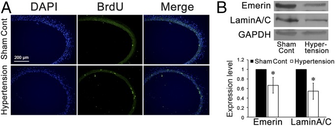
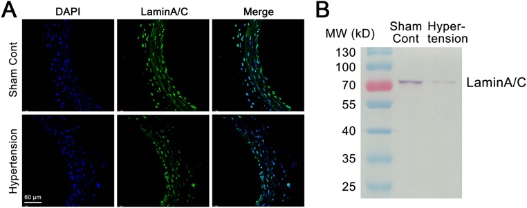
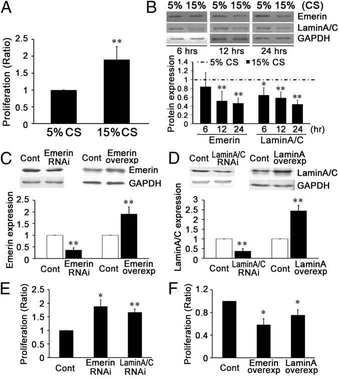
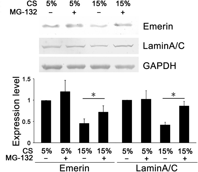
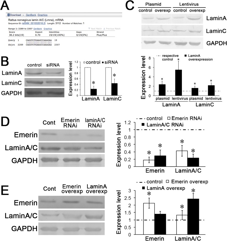
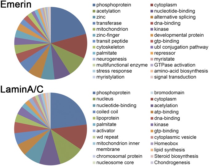
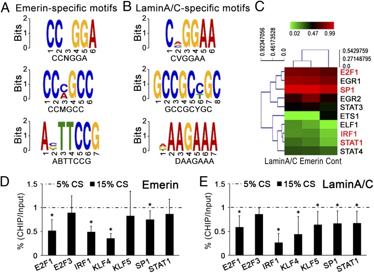
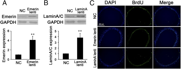
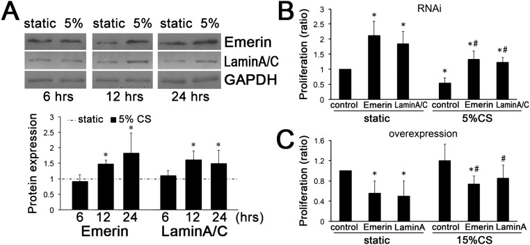
Cyclic stretch is an important inducer of vascular smooth muscle cell (VSMC) proliferation, which is crucial in vascular remodeling during hypertension. However, the molecular mechanism remains unclear. We studied the effects of emerin and lamin A/C, two important nuclear envelope proteins, on VSMC proliferation in hypertension and the underlying mechano-mechanisms. In common carotid artery of hypertensive rats in vivo and in cultured cells subjected to high (15%) cyclic stretch in vitro, VSMC proliferation was increased significantly, and the expression of emerin and lamin A/C was repressed compared with normotensive or normal (5%) cyclic stretch controls. Using targeted siRNA to mimic the repressed expression of emerin or lamin A/C induced by 15% stretch, we found that VSMC proliferation was enhanced under static and 5%-stretch conditions. Overexpression of emerin or lamin A/C reversed VSMC proliferation induced by 15% stretch. Hence, emerin and lamin A/C play critical roles in suppressing VSMC hyperproliferation induced by hyperstretch. ChIP-on-chip and MOTIF analyses showed that the DNAs binding with emerin contain three transcription factor motifs: CCNGGA, CCMGCC, and ABTTCCG; DNAs binding with lamin A/C contain the motifs CVGGAA, GCCGCYGC, and DAAGAAA. Protein/DNA array proved that altered emerin or lamin A/C expression modulated the activation of various transcription factors. Furthermore, accelerating local expression of emerin or lamin A/C reversed cell proliferation in the carotid artery of hypertensive rats in vivo. Our findings establish the pathogenetic role of emerin and lamin A/C repression in stretch-induced VSMC proliferation and suggest mechanobiological mechanism underlying this process that involves the sequence-specific binding of emerin and lamin A/C to specific transcription factor motifs.

Keywords: emerin; laminA/C; mechanobiology; specific-binding sequence; transcription factors.


Conflict of interest statement

The authors declare no conflict of interest.

Figures

Fig. 1.
Fig. 1.
In situ proliferation and expression of emerin and lamin A/C in the common carotid artery of hypertensive rats and sham-treated controls. (A) Immunofluorescence staining against BrdU revealed that after 1 wk of abdominal aorta coarctation, VSMC proliferation in the media of common carotid arteries increased markedly. (B) The expression of emerin and of lamin A/C was repressed significantly in the common carotid arteries of the aorta-coarctation–induced hypertensive rats. GAPDH was used for normalization. Values are expressed as mean ± SD. *P < 0.05 vs. the sham-treated control (n = 5).
Fig. S1.
Fig. S1.
The expression of lamin A/C in the common carotid arteries of hypertensive rats was repressed in comparison with the expression in sham controls. (A) Immunofluorescence staining of lamin A/C in the common carotid artery is higher in a normal control rat than in a hypertensive rat. (B) Entire immunoblots of lamin A/C in the common carotid arteries of aorta-coarctation hypertensive rats and sham-operated controls.
Fig. 2.
Fig. 2.
Cyclic stretch (CS) modulated the expression of NE proteins emerin and lamin A/C, which participate in the stretch-induced proliferation of VSMCs in vitro. (A) A 15% cyclic stretch increased VSMC proliferation in comparison with a 5% cyclic stretch. (B) A 15% cyclic stretch decreased the expression of emerin and lamin A/C in comparison with a 5% cyclic stretch. (C) Under static conditions, target siRNA transfection significantly decreased and plasmid transfection significantly increased the expression of emerin. (D) Under static conditions, target siRNA transfection significantly decreased and plasmid transfections significantly increased the expression of lamin A/C. (E) Under static conditions, specific RNAi of both emerin and lamin A/C increased VSMC proliferation. (F) Up-regulated expression of emerin and lamin A/C decreased VSMC proliferation. For Western blots, GAPDH was used for normalization. Values are expressed as mean ± SD. *P < 0.05, **P < 0.01 vs. the respective control (Cont) (n = 5).
Fig. S2.
Fig. S2.
MG-132(R), a proteasome inhibitor, increased expression of emerin and lamin A/C in VSMCs subjected to 15% cyclic stretch for 24 h. GAPDH was used for normalization. Values are expressed as mean ± SD. *P < 0.05 vs. the respective control (n = 4). CS, cyclic stretch.
Fig. S3.
Fig. S3.
The expressions of emerin, lamin A, and lamin C are correlated with one other. (A) The siRNA (CAGG UCUG AAGC CAAA GAAT T) targets on LMNA, which is the template of both lamin A and lamin C. (B) Transfection of target siRNA to lamin A/C modulated the expression of both lamin A and lamin C. GAPDH was used for normalization. Values are expressed as mean ± SD. *P < 0.05 vs. the respective control (n = 4). (C) Overexpression of lamin A with either plasmid or lentivirus markedly increased the expression of both lamin A and lamin C. GAPDH was used for normalization. Values are expressed as mean ± SD. *P < 0.05 vs. the respective control (n = 4). (D) After transfection of emerin siRNA for 48 h, the expression of lamin A/C was decreased; conversely, lamin A/C siRNA also decreased the expression of emerin. (E) Emerin overexpression increased the expression of lamin A/C; lamin A overexpression increased the expression of lamin A/C but had no significant effect on the expression of emerin. GAPDH was used for normalization. Values are expressed as mean ± SD. *P < 0.05 vs. the respective control (n = 6).
Fig. 3.
Fig. 3.
Cyclic stretch modulates the proliferation of VSMCs via NE proteins in vitro. (A) The expression of emerin or lamin A/C was repressed by target siRNA transfection during the application of 5% cyclic stretch. (B) Emerin- or lamin A/C-target siRNA transfection increased the proliferation of VSMCs. (C) The expression of emerin and of lamin A/C was increased by plasmid transfection. (D) Transfection with plasmid overexpressing emerin and lamin A decreased VSMC proliferation under 15% cyclic stretch. For Western blots, GAPDH was used for normalization. Values are expressed as mean ± SD. *P < 0.05, **P < 0.01 vs. the respective control (n = 6).
Fig. S4.
Fig. S4.
Entire immunoblots of lamin A/C in cultured VSMCs and common carotid arteries. (A) Entire immunoblots of lamin A/C in VSMCs transfected with siRNA during the application of 5% cyclic stretch. (B) Entire immunoblots of lamin A/C in VSMCs transfected with overexpression plasmid of lamin A during the application of 15% cyclic stretch. (C) Entire immunoblots of lamin A/C in the common carotid arteries of hypertensive rats locally injected with lentiviruses of lamin A or the respective control. NC, negative control.
Fig. 4.
Fig. 4.
GO annotations show the primary functional categories of the DNA segments immunoprecipitated with emerin or lamin A/C (Table S2).
Fig. 5.
Fig. 5.
Motifs of transcription factors enriched in DNA segments binding with emerin or lamin A/C. (A) The specific motifs in the DNA segments immunoprecipitated with emerin. (B) The specific motifs in the DNA segments immunoprecipitated with lamin A/C. (C) Emerin or lamin A/C-target siRNA modulated the activations of transcription factors in Table S4. The red font indicates the factors involved in the proliferation of smooth muscle cells. (D and E) After immunoprecipitation with emerin (D) or lamin A/C (E), qPCR detected the ChIP levels of promoter regions of transcription factors related with “proliferation of (vascular) smooth muscle cells” in Table S4 after cyclic stretch was applied for 24 h. Values are expressed as mean ± SD (n = 4).
Fig. 6.
Fig. 6.
(A and B) Local injection of emerin (A) or lamin A (B) lentiviruses (lenti) significantly increased the expression of emerin or lamin A/C in common carotid arteries the hypertensive rats. (C) Immunofluorescence staining against BrdU revealed that locally injected lentiviruses of emerin or lamin A remarkably decreased VSMC proliferation in the media of common carotid arteries of hypertensive rats. For Western blot, GAPDH was used for normalization. Values are expressed as mean ± SD. **P < 0.01 vs. the negative control (NC) (n = 6).
Fig. S5.
Fig. S5.
Emerin and lamin A/C negatively regulate VSMC proliferation under different conditions (static control, 5% cyclic stretch, and 15% cyclic stretch). (A) Emerin and lamin A/C expression was increased after 5% cyclic stretch (CS) applied for 12 h and 24 h, compared with static control. GAPDH was used for normalization. Values are expressed as mean ± SD; *P < 0.05 vs. the static control (n = 5). (B and C) Emerin and lamin A/C were repressed by RNAi (B) or overexpressed by plasmid transfection (C). Then VSMCs were subjected to 5% and 15% cyclic stretch. The results indicate that both emerin and lamin A/C exert an inhibitory effect on cell proliferation under stretch conditions. *P < 0.05 vs. the static control, #P < 0.05 vs. the respective control under cyclic stretch application (n = 4).

References

    1. Halka AT, et al. The effects of stretch on vascular smooth muscle cell phenotype in vitro. Cardiovasc Pathol. 2008;17(2):98–102. - PubMed
    1. Haga JH, Li YS, Chien S. Molecular basis of the effects of mechanical stretch on vascular smooth muscle cells. J Biomech. 2007;40(5):947–960. - PubMed
    1. Morrow D, et al. Cyclic strain inhibits Notch receptor signaling in vascular smooth muscle cells in vitro. Circ Res. 2005;96(5):567–575. - PubMed
    1. Qi YX, et al. Cyclic strain modulates migration and proliferation of vascular smooth muscle cells via Rho-GDIalpha, Rac1, and p38 pathway. J Cell Biochem. 2010;109(5):906–914. - PubMed
    1. Butler PJ, Tsou TC, Li JY, Usami S, Chien S. Rate sensitivity of shear-induced changes in the lateral diffusion of endothelial cell membrane lipids: A role for membrane perturbation in shear-induced MAPK activation. FASEB J. 2002;16(2):216–218. - PubMed

Publication types