. 2016 May 10;113(19):5435-40.
doi: 10.1073/pnas.1604453113. Epub 2016 Apr 25.

Tryptophan-2,3-dioxygenase (TDO) inhibition ameliorates neurodegeneration by modulation of kynurenine pathway metabolites

Affiliations

Tryptophan-2,3-dioxygenase (TDO) inhibition ameliorates neurodegeneration by modulation of kynurenine pathway metabolites

Carlo Breda et al. Proc Natl Acad Sci U S A. .

Abstract

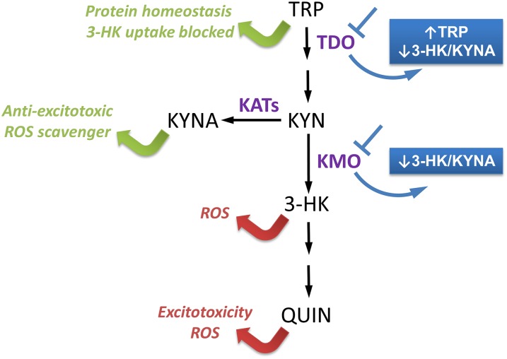
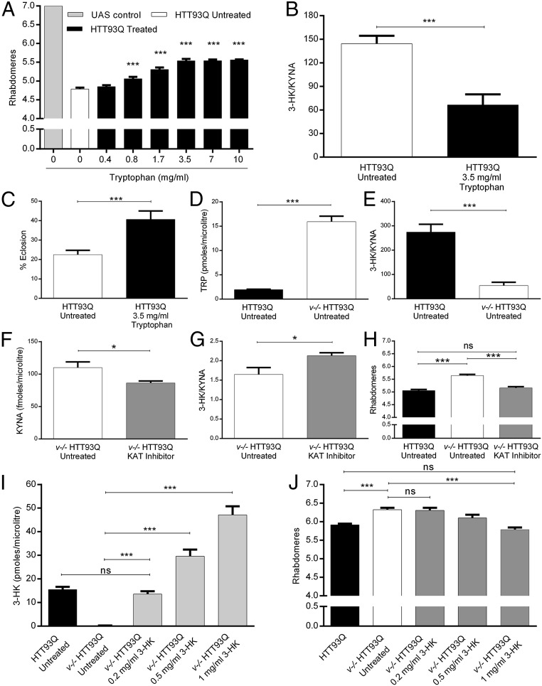
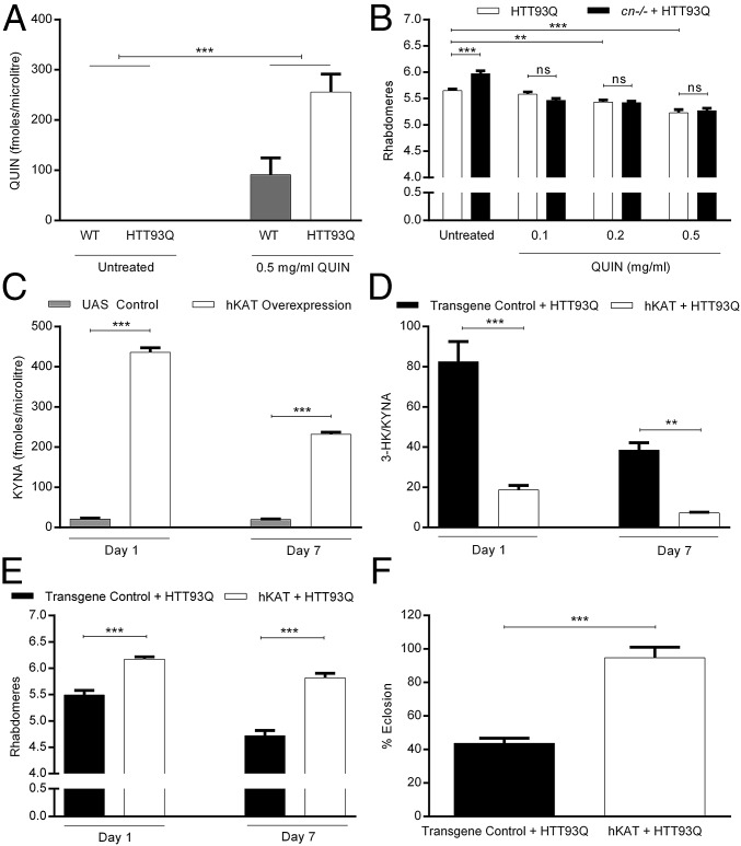
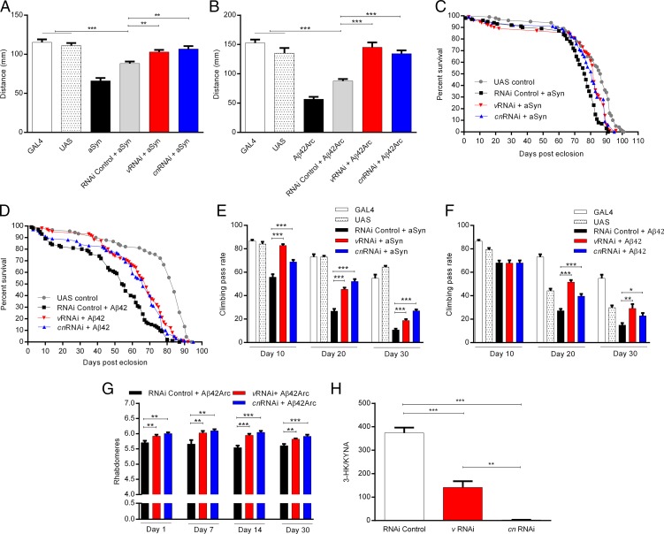
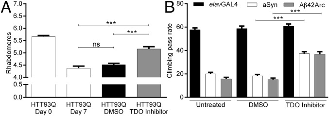
Metabolites of the kynurenine pathway (KP) of tryptophan (TRP) degradation have been closely linked to the pathogenesis of several neurodegenerative disorders. Recent work has highlighted the therapeutic potential of inhibiting two critical regulatory enzymes in this pathway-kynurenine-3-monooxygenase (KMO) and tryptophan-2,3-dioxygenase (TDO). Much evidence indicates that the efficacy of KMO inhibition arises from normalizing an imbalance between neurotoxic [3-hydroxykynurenine (3-HK); quinolinic acid (QUIN)] and neuroprotective [kynurenic acid (KYNA)] KP metabolites. However, it is not clear if TDO inhibition is protective via a similar mechanism or if this is instead due to increased levels of TRP-the substrate of TDO. Here, we find that increased levels of KYNA relative to 3-HK are likely central to the protection conferred by TDO inhibition in a fruit fly model of Huntington's disease and that TRP treatment strongly reduces neurodegeneration by shifting KP flux toward KYNA synthesis. In fly models of Alzheimer's and Parkinson's disease, we provide genetic evidence that inhibition of TDO or KMO improves locomotor performance and ameliorates shortened life span, as well as reducing neurodegeneration in Alzheimer's model flies. Critically, we find that treatment with a chemical TDO inhibitor is robustly protective in these models. Consequently, our work strongly supports targeting of the KP as a potential treatment strategy for several major neurodegenerative disorders and suggests that alterations in the levels of neuroactive KP metabolites could underlie several therapeutic benefits.

Keywords: Alzheimer’s disease; KMO; Parkinson’s disease; TDO; neurodegeneration.


Conflict of interest statement

The authors declare no conflict of interest.

Figures

Fig. 1.
Fig. 1.
Consequences of KP manipulation. KP metabolites and enzymatic steps are indicated in black, whereas the key KP enzymes TDO, KMO, and KATs are indicated in purple. The metabolites 3-HK and QUIN are neurotoxic (as indicated by red arrows), whereas KYNA and TRP are neuroprotective (as indicated by green arrows). Inhibition of TDO results in increased TRP levels, and either TDO or KMO inhibition leads to a reduction in the 3-HK/KYNA ratio (highlighted in blue). The enzyme 3-hydroxyanthranilic acid dioxygenase is not present in flies, and thus QUIN is not synthesized.
Fig. 2.
Fig. 2.
TRP feeding ameliorates HTT93Q toxicity in fruit flies. (A) Rhabdomere quantification of HD flies treated with different concentrations of TRP during development. TRP concentrations higher than 0.4 mg/mL significantly ameliorate rhabdomere neurodegeneration. n = 6–13 per condition, ***P < 0.001. (B) The 3-HK/KYNA ratio is reduced in TRP-fed HTT93Q flies. n = 5–6 per condition, ***P < 0.001. (C) TRP feeding rescues HTT93Q-dependent eclosion defects. Untreated HD flies: n = 938; TRP-treated HD flies: n = 728, ***P < 0.001. (D) TRP levels are significantly increased in v−/− HTT93Q flies compared with HTT93Q flies. n = 5 per condition, ***P < 0.001. (E) 3-HK/KYNA levels are reduced in v−/− HTT93Q compared with HTT93Q flies. n = 5 flies per condition, ***P < 0.001. (F) Treatment with the KAT inhibitor AOAA (100 μM in the food) reduces the level of KYNA in v−/− HTT93Q flies. n = 5 per condition. *P < 0.05. (G) AOAA treatment leads to a reduction in the 3-HK/KYNA ratio. n = 5 per condition, *P < 0.05. (H) KAT inhibition abrogates the neuroprotection conferred by the v mutation. n = 12–14 per condition, ***P < 0.001; ns, not significant. (I) Feeding of 3-HK leads to increased levels of 3-HK in v−/− HTT93Q flies. n = 5–6 per treatment. ***P < 0.001; ns, not significant. (J) Supplementation of 3-HK in the food of v−/− HTT93Q flies reduces neuroprotection compared with untreated HD flies. n = 8–12 per condition, ***P < 0.001; ns, not significant. Data are the mean ± SEM (one-way ANOVA with Newman–Keuls post hoc test).
Fig. 3.
Fig. 3.
QUIN exacerbates neurodegeneration in HD flies and overexpression of hKAT is neuroprotective via increased KYNA levels. (A) QUIN levels in WT and HTT93Q-expressing flies. QUIN is detected in flies fed with 0.5 mg/mL of QUIN, but was not measurable in untreated flies. n = 3–5 flies per treatment, ***P < 0.001. (B) HTT93Q and cn−/− HTT93Q flies fed QUIN exhibit increased rhabdomere degeneration compared with untreated flies. Neuroprotection conferred by the cn mutation is abolished by QUIN feeding. n = 11–12 per treatment, **P < 0.01, ***P < 0.001. (C) Panneuronal overexpression of hKAT in a WT background causes an increase in KYNA production compared with controls at both posteclosion ages tested. n = 3–5 per genotype, ***P < 0.001. (D) HTT93Q flies with panneuronal overexpression of hKAT show a significant reduction in the 3-HK/KYNA ratio. The transgene control used in this experiment was a transgenic Drosophila line expressing an empty pJFRC2 vector. n = 4–5 per condition, **P < 0.01, ***P < 0.001. (E) Overexpression of hKAT is neuroprotective in HTT93Q flies at both posteclosion ages tested. n = 9–13 flies per condition, ***P < 0.001. (F) Overexpression of hKAT ameliorates the eclosion phenotype observed in HTT93Q flies. Transgene control + Htt93Q flies: n = 1084; hKAT + Htt93Q flies: n = 1,010, ***P < 0.001; ns, not significant. Data are the mean ± SEM (one-way ANOVA with Newman–Keuls post hoc test).
Fig. 4.
Fig. 4.
v and cn down-regulation ameliorates PD- and AD-related impairments in Drosophila. Expression of aSyn (A) or Aβ42ARC (B) in motorneurons using the c164GAL4 driver reduces the distance crawled by third instar larvae. The silencing of v or cn significantly ameliorates these locomotor defects. n = 20 larvae per genotype. **P < 0.01 and ***P < 0.001. Panneuronal expression of aSyn (C) or Aβ42 (D) reduces average life span, which is reversed by v and cn silencing. n = 100 per genotype. Median survival in days for aSyn experiments: UAS control = 86; RNAi control + aSyn = 76; vRNAi + aSyn = 82; cnRNAi + aSyn = 81. Median survival in days for Aβ42 experiments: UAS control = 84; RNAi control + Aβ42 = 54; vRNAi + Aβ42 = 68; cnRNAi + Aβ42 = 66. (E and F) Mean climbing pass rate at different posteclosion ages for flies expressing aSyn or Aβ42 panneuronally. Both aSyn (E) and Aβ42 (F) reduce climbing performance, and the effects are reversed by down-regulation of v and cn. n = 50–60 per genotype and condition. *P < 0.05, **P < 0.01, and ***P < 0.001. Panneuronal expression of Aβ42Arc reduces mean rhabdomeres per ommatidium (G); v and cn silencing protects rhabdomere degeneration at all posteclosion ages tested. n = 7–11 per condition. **P < 0.01 and ***P < 0.001. (H) The 3-HK/KYNA ratio is decreased in flies with RNAi down-regulation of v and cn. n = 5 per genotype. **P < 0.01 and ***P < 0.001. Data are the mean ± SEM (one-way ANOVA with Newman–Keuls post hoc test).
Fig. 5.
Fig. 5.
Pharmacological inhibition of TDO is neuroprotective in HD, PD, and AD flies. (A) Reduced neurodegeneration in HTT93Q flies treated with the TDO inhibitor 680C91 (100 µM) 7 d posteclosion. n = 8–17 per condition. (B) aSyn and Aβ42Arc flies treated with 680C91 (100 µM) display improved climbing compared with controls. n = 50–60 per genotype. DMSO, dimethyl sulfoxide. ***P < 0.001; ns, not significant. Data are the mean ± SEM (one-way ANOVA with Newman–Keuls post hoc test).

Similar articles

Cited by

References

    1. Amaral M, Outeiro TF, Scrutton NS, Giorgini F. The causative role and therapeutic potential of the kynurenine pathway in neurodegenerative disease. J Mol Med (Berl) 2013;91(6):705–713. - PubMed
    1. Thevandavakkam MA, Schwarcz R, Muchowski PJ, Giorgini F. Targeting kynurenine 3-monooxygenase (KMO): Implications for therapy in Huntington’s disease. CNS Neurol Disord Drug Targets. 2010;9(6):791–800. - PubMed
    1. Stone TW, Perkins MN. Quinolinic acid: A potent endogenous excitant at amino acid receptors in CNS. Eur J Pharmacol. 1981;72(4):411–412. - PubMed
    1. Schwarcz R, Whetsell WO, Jr, Mangano RM. Quinolinic acid: An endogenous metabolite that produces axon-sparing lesions in rat brain. Science. 1983;219(4582):316–318. - PubMed
    1. Okuda S, Nishiyama N, Saito H, Katsuki H. Hydrogen peroxide-mediated neuronal cell death induced by an endogenous neurotoxin, 3-hydroxykynurenine. Proc Natl Acad Sci USA. 1996;93(22):12553–12558. - PMC - PubMed

Publication types

MeSH terms