. 2016 May 10;113(19):5447-52.
doi: 10.1073/pnas.1522458113. Epub 2016 Apr 25.

Recruitment of the NineTeen Complex to the activated spliceosome requires AtPRMT5

Affiliations

Recruitment of the NineTeen Complex to the activated spliceosome requires AtPRMT5

Xian Deng et al. Proc Natl Acad Sci U S A. .

Erratum in

Abstract

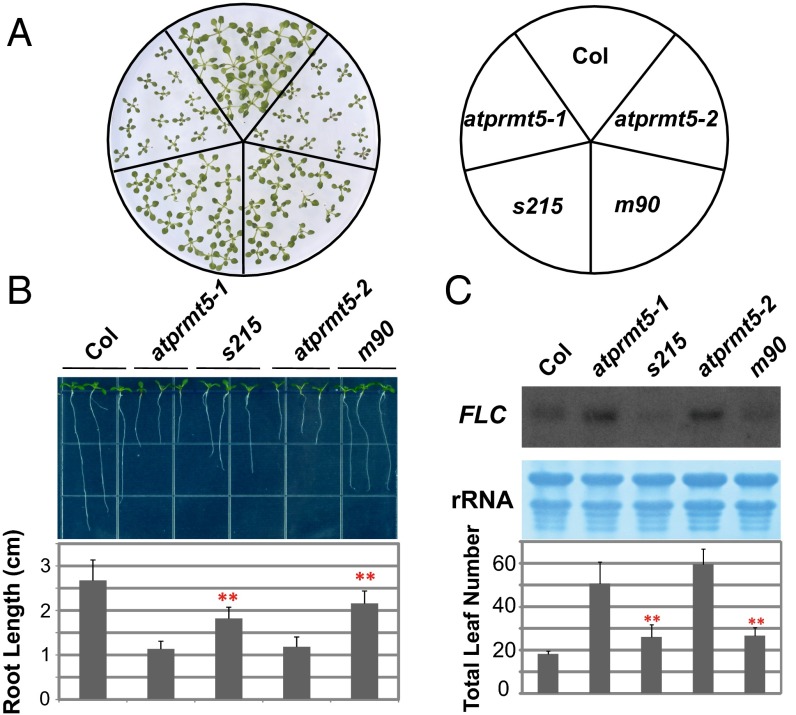
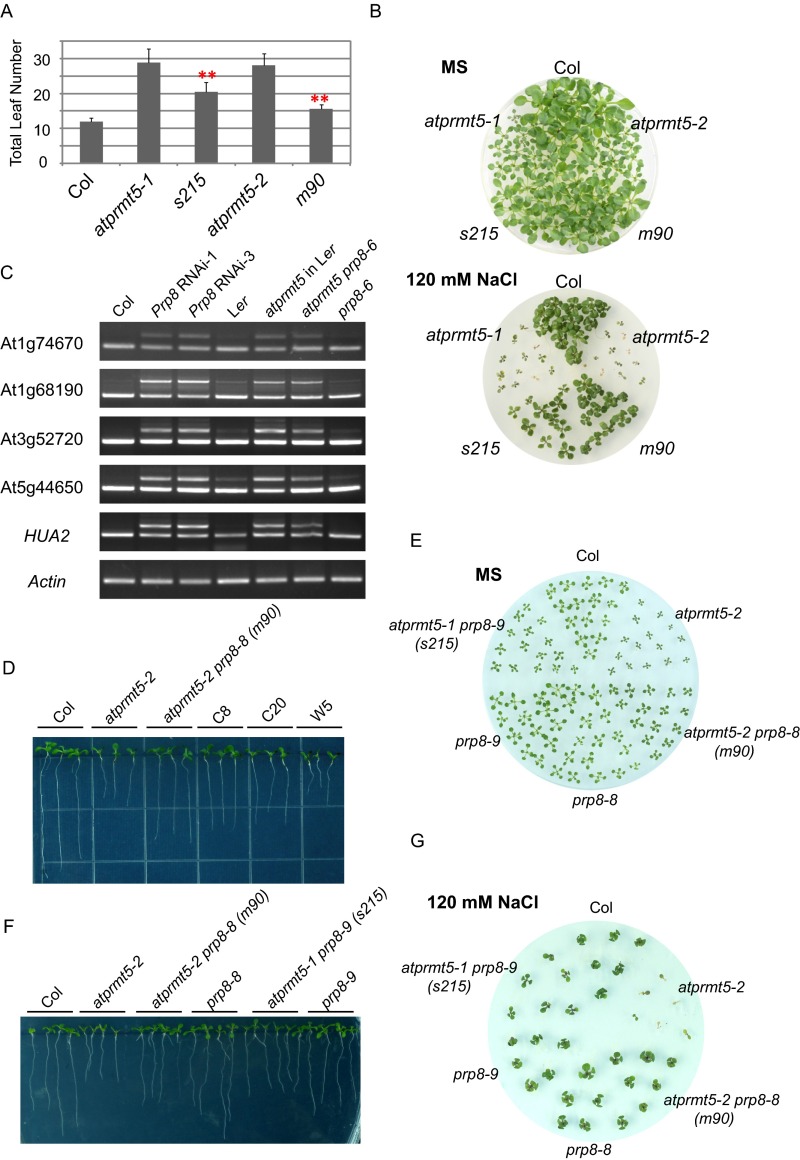
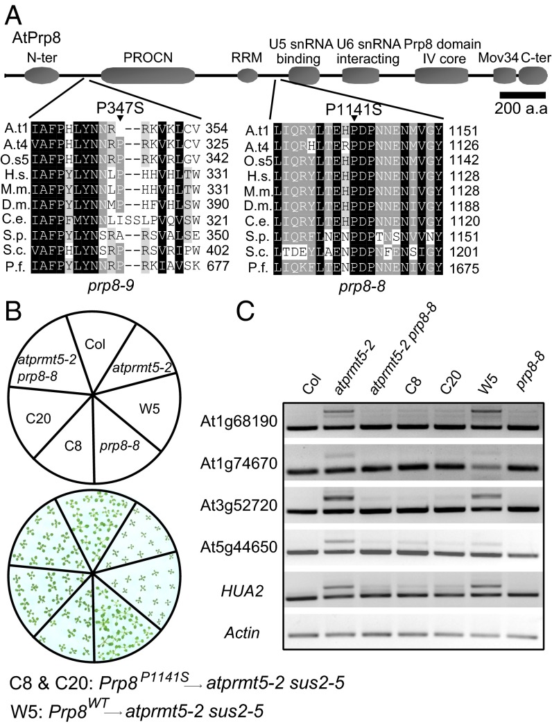
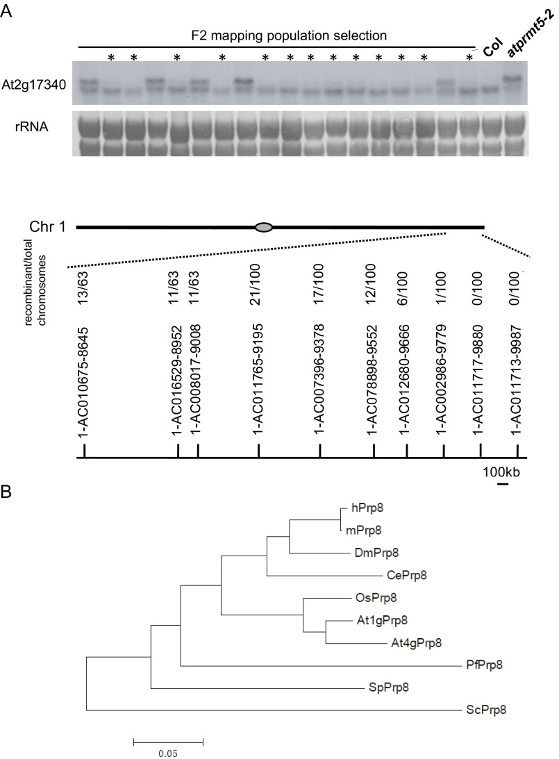
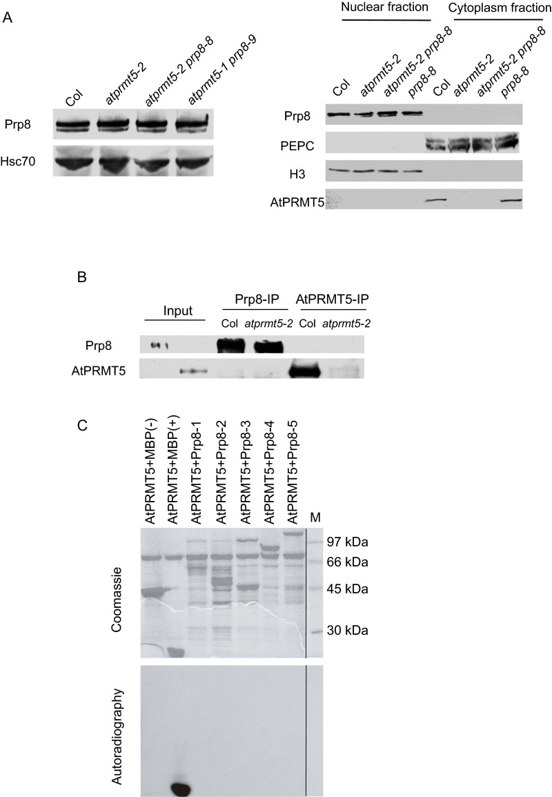
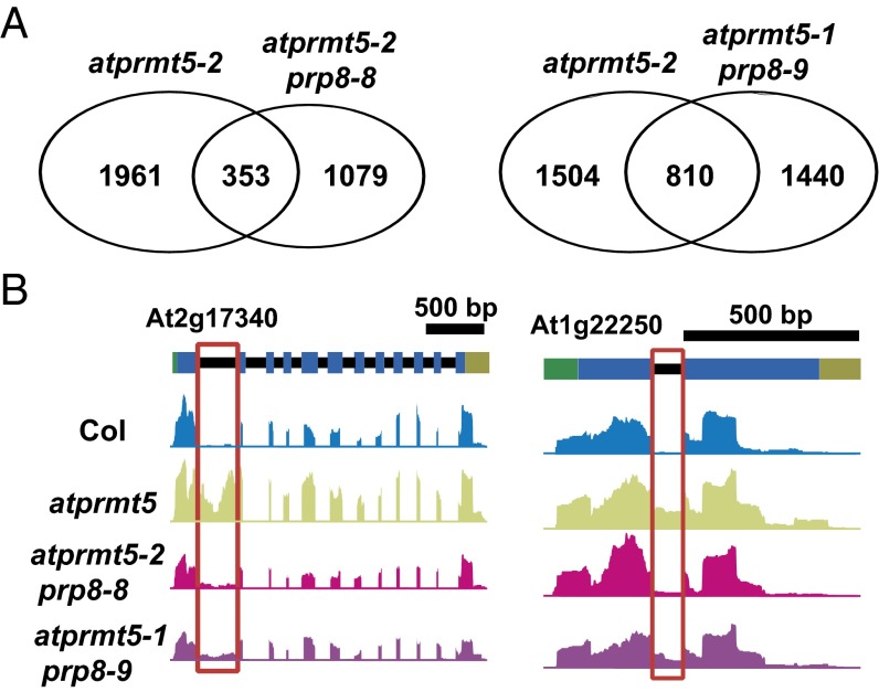
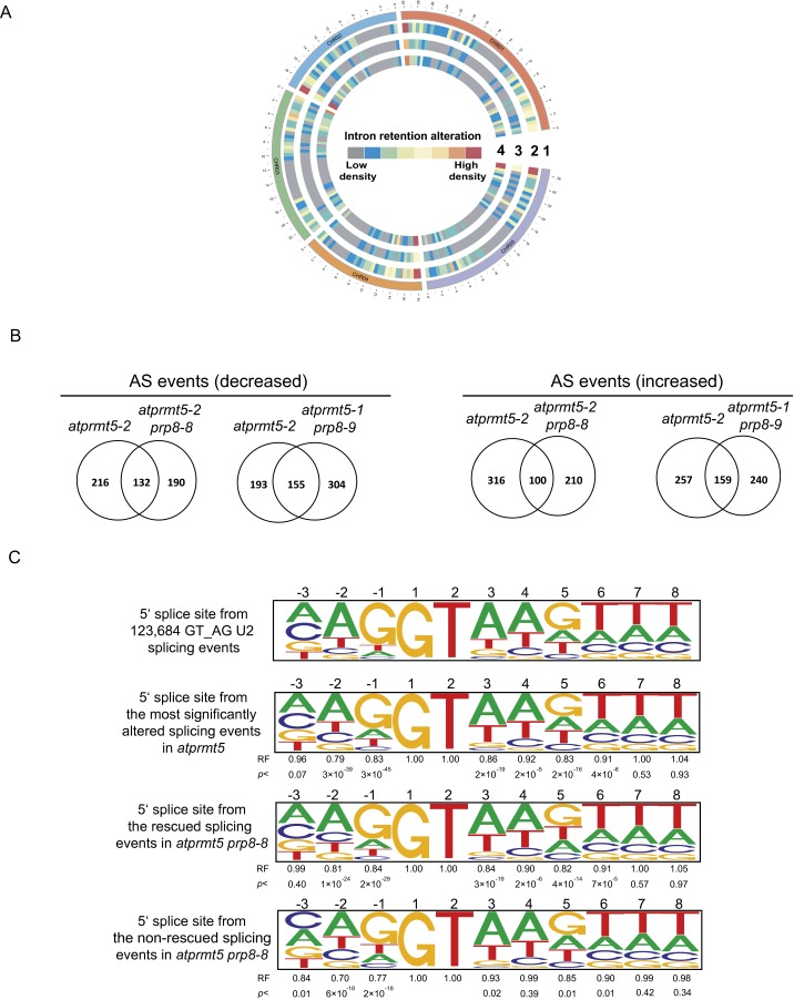
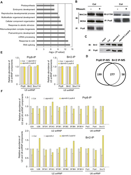
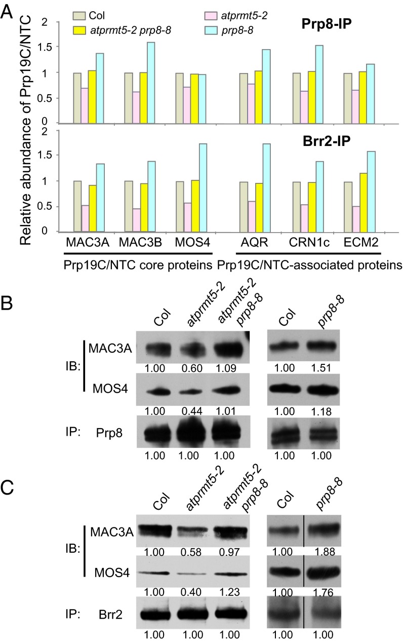
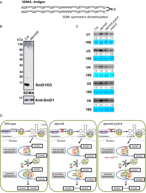
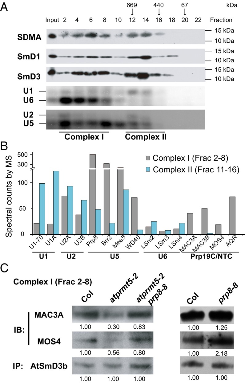
Protein arginine methylation, catalyzed by protein arginine methyltransferases (PRMTs), is involved in a multitude of biological processes in eukaryotes. Symmetric arginine dimethylation mediated by PRMT5 modulates constitutive and alternative pre-mRNA splicing of diverse genes to regulate normal growth and development in multiple species; however, the underlying molecular mechanism remains largely unknown. A genetic screen for suppressors of an Arabidopsis symmetric arginine dimethyltransferase mutant, atprmt5, identified two gain-of-function alleles of pre-mRNA processing factor 8 gene (prp8-8 and prp8-9), the highly conserved core component of the U5 small nuclear ribonucleoprotein (snRNP) and the spliceosome. These two atprmt5 prp8 double mutants showed suppression of the developmental and splicing alterations of atprmt5 mutants. In atprmt5 mutants, the NineTeen complex failed to be assembled into the U5 snRNP to form an activated spliceosome; this phenotype was restored in the atprmt5 prp8-8 double mutants. We also found that loss of symmetric arginine dimethylation of Sm proteins prevents recruitment of the NineTeen complex and initiation of spliceosome activation. Together, our findings demonstrate that symmetric arginine dimethylation has important functions in spliceosome assembly and activation, and uncover a key molecular mechanism for arginine methylation in pre-mRNA splicing that impacts diverse developmental processes.

Keywords: AtPRMT5; Prp19C/NTC; arginine methylation; pre-mRNA splicing; protein arginine methyltransferase.


Conflict of interest statement

The authors declare no conflict of interest.

Figures

Fig. 1.
Fig. 1.
atprmt5 suppressors partially rescue the pleiotropic developmental defects of atprmt5 mutants. (A) Rescued growth retardation of young seedling leaves at 12 d. (B) Rescued primary roots of s215 and m90 at 9 d. Data are shown as means ± SD (n = 20). Two-sided Student t test between atprmt5 and the suppressors was performed (**P < 0.01). (C) Rescued FLC expression and flowering time of s215 and m90. (Upper) The total RNAs from 12-d-old seedlings of Col, atprmt5, s215, and m90 plants were probed by RNA blot with the full-length coding sequence of FLC. rRNAs were used as a loading control. (Lower) Flowering time was assessed by total leaf number after plants stopped producing new leaves when plants were grown at 23 °C under long day conditions. Data are shown as means ± SD (n = 30). Two-sided Student t test between atprmt5 and the suppressors was performed (**P < 0.01).
Fig. S1.
Fig. S1.
Phenotypic and molecular analysis of atprmt5 suppressors. (A) Flowering time of Col, atprmt5 mutants, s215, and m90, assessed by total leaf number after plants stop producing new leaves. Plants were subjected to vernalization treatment at 4 °C for 6 wk and then transferred to 23 °C and grown under long day conditions. Data are shown as means ± SD (n = 20). Two-sided Student t test between atprmt5 and the suppressors was performed (**P < 0.01). (B) Comparison of the survival rate of the Col, atprmt5 mutants, s215, and m90 plants grown on MS medium containing 120 mM NaCl. Photographs were taken 25 d after seedling transfer to the treatment medium. (C) RT-PCR analysis for Prp8 RNAi lines in atprmt5 background and atprmt5-2 prp8-6 double mutants (in Ler background). (D) Rescued primary roots of the transformation lines of CDS Prp8 clone with P1141S mutation in atprmt5-2 sus2-5 double mutants background at 9 d. (E–G) Phenotypic analysis of the young seedling leaves at 12 d (E), the primary roots at 9 d (F), and the survival rate on MS medium containing 120 mM NaCl (G) for prp8-8 and prp8-9 mutants.
Fig. 2.
Fig. 2.
Point mutations in Prp8 are responsible for suppressors of atprmt5 mutants. (A) Sequence alignment of the conserved Prp8 containing the P347S and P1141S mutations in prp8-9 and prp8-8, respectively. Abbreviations for species are as follows: Arabidopsis thaliana (At), Oryza sativa (Os), Homo sapiens (Hs), Mus musculus (Mm), Drosophila melanogaster (Dm), Caenorhabditis elegans (Ce), Schizosaccharomyces pombe (Sp), Saccharomyces cerevisiae (Sc), and Plasmodium falciparum (Pf). The point mutation sites are labeled by black triangles. (B and C) Rescued growth retardation of young seedling leaves at 12 d (B) and intron retention events (C) by a transformation of CDS Prp8 clone with P1141S mutation in atprmt5-2 sus2-5 double mutants background. C8 and C20 are two individual transgenic lines containing P1141S construct of Prp8. W5 is the control transgenic line containing WT Prp8 construct.
Fig. S2.
Fig. S2.
Low-resolution mapping and resequencing of the m90 mutation indicate it has a lesion in At1g80070. (A) (Upper) RNA blot analysis of the splicing patterns of At2g17340 in the F2 mapping population of m90, with rRNA as the loading control. The lanes marked with asterisks represent the population used for low-resolution mapping. The F2 population segregated into WT and mutant phenotypes at a ratio of 3:1, indicating that the mutant phenotype is caused by a single recessive mutation; (Lower) Bulked segregant analysis maps atprmt5-2 prp8-8 to between 1-AC002986-9779 and 1-AC011713-9987 on chromosome 1. (B) Phylogenic analysis of Prp8 from yeast, fly, mouse, worm, human, Arabidopsis, and rice. The species for each protein is designated by the following prefixes: Arabidopsis thaliana (At), Oryza sativa (Os), Homo sapiens (Hs), Mus musculus (Mm), Drosophila melanogaster (Dm), Caenorhabditis elegans (Ce), Schizosaccharomyces pombe (Sp), Saccharomyces cerevisiae (Sc), and Plasmodium falciparum (Pf).
Fig. S3.
Fig. S3.
Point mutations in Prp8 do not destabilize protein levels, and AtPRMT5 does not interact with and methylate Prp8. (A) Analysis of Prp8 protein levels and nucleus/cytoplasm distribution by Western blotting using anti-Prp8 polyclonal antibody, with Hsc70, PEPC, and H3 as loading controls for total, cytoplasm, and nucleus, respectively. (B) AtPRMT5 does not interact with Prp8 in vivo. AtPRMT5 and Prp8 immunoprecipitated by anti-AtPRMT5 and anti-Prp8 polyclonal antibodies from Col and atprmt5 mutants were immunoblotted with anti-Prp8 and anti-AtPRMT5 antibodies, respectively. (C) AtPRMT5 does not methylate Prp8 in vitro. Full length of Prp8 is divided into five fragments (–5), and each of the fragments is subjected to the methyltransferase activity assay with AtPRMT5. The dividing lines indicate noncontiguous images between samples and the protein marker.
Fig. 3.
Fig. 3.
atprmt5 suppressors partially rescue the splicing alterations of atprmt5 mutants. (A) Overlapping analysis of significantly altered constitutive splicing events between atprmt5-2 and the suppressors. (B) Two examples of intron retention events rescued in atprmt5-1 prp8-9 and atprmt5-2 prp8-8. Annotated gene structures are shown (Upper), with thick lines representing exons and thin lines representing introns. Wiggle plots representing the normalized reads coverage in a logarithmic scale (log2) are shown in blue for Col, in green for atprmt5 mutants, in pink for atprmt5-2 prp8-8, and in purple for atprmt5-1 prp8-9. The red frames indicate the retained introns rescued in atprmt5-1 prp8-9 and atprmt5-2 prp8-8.
Fig. S4.
Fig. S4.
Impact of atprmt5-2, atprmt5 prp8-8, and atprmt5 prp8-9 on genome-wide constitutive splicing and alternative splicing. (A) The global splicing defects of atprmt5 mutants are partially suppressed in atprmt5-1 prp8-9 and atprmt5-2 prp8-8 plants. The heatmap view of the density of pre-mRNA splicing defects in a 600-kb window in atprmt5-2 (circle 2), atprmt5-2 prp8-8 (circle 3), and atprmt5-1 prp8-9 (circle 4). Circle 1 represents the chromosome. (B) Overlap between significantly altered alternative splicing events. (C) Bioinformatic analysis of donor splice-site sequences. Pictograms showing the frequency distribution of nucleotides at the 5′ splice site of 123,684 GT_AG_U2 Arabidopsis introns, the most significantly intron retention events whose splicing were altered simultaneously in atprmt5 and the significantly rescued and nonrescued intron retention events in atprmt5 prp8-8. The representation factor (RF) is the frequency of interest divided by the total frequency. For each RF, a P value was calculated using the hypergeometric test.
Fig. S5.
Fig. S5.
Prp8 interacts with Brr2 and share similar interaction proteins. (A) Gene ontology terms enriched in genes associated with Prp8. (B) The interactions between Prp8 and Prp19C/NTC are independent of RNA (Left), whereas the interaction between Prp8 and U1 snRNP (U1-70K) is partially dependent on RNA (Right). (C) Prp8 immunoprecipitated by anti-Prp8 antibody was immunoblotted with anti-Brr2 antibody. (D) Venn diagram of numbers of Prp8-associated and Brr2-associated proteins, as identified by MS. (E and F) The association of Prp8 (and Brr2) within U5 snRNP (E) and U2 and U6 snRNPs (F) in Col, atprmt5-2, and atprmt5-2 prp8-8.
Fig. 4.
Fig. 4.
The recruitment of Prp19C/NTC to the spliceosome is impaired in atprmt5 mutants and restored in atprmt5-2 prp8-8. (A) The association of Prp19C/NTC and Prp8 (and Brr2) decreased in atprmt5 mutants and was rescued in atprmt5-2 prp8-8 mutants. Nuclear extracts from Col, atprmt5-2, atprmt5-2 prp8-8, and prp8-8 were immunoprecipitated by anti-Prp8 (Upper) and anti-Brr2 (Lower) polyclonal antibodies, and the interacting proteins were identified by MS. The spectral counts of Prp19C/NTC from atprmt5-2, atprmt5-2 prp8-8, and prp8-8 were divided by that from Col, and the ratios are shown in the histogram. (B and C) Prp8- or Brr2-associated proteins were identified in Col, atprmt5, atprmt5-2 prp8-8, and prp8-8 by MS. Prp8 (B) and Brr2 (C) were immunoprecipitated by anti-Prp8 and anti-Brr2 antibodies, respectively, and immunoblotting was performed with anti-MAC3A and anti-MOS4 antibodies for Prp19C/NTC. The dividing lines in C, Right indicate noncontiguous images between Col and prp8-8.
Fig. S6.
Fig. S6.
Preparation of anti-SDMA antibody, U snRNAs analysis, and working model. (A) Low complex GAR motifs harboring symmetric dimethylarginines (SDMA). (B) Total proteins extracted from Col and atprmt5 mutants were immunoblotted with anti-SDMA and anti-SmD1 antibodies. (C) Levels of U1, U2, U4, U5, and U6 snRNAs are increased in atprmt5 mutants and restored in atprmt5 prp8-8. 18S rRNAs were used as a loading control. (D) Model for the role of AtPRMT5 in Prp19C/NTC complex recruitment to the spliceosome during pre-mRNA splicing.
Fig. 5.
Fig. 5.
Decreased recruitment of Prp19C/NTC to the spliceosome in atprmt5 mutants. (A) The elution profiles of symmetric dimethylated AtSmD1 and AtSmD3 proteins. Cell lysates from Col were fractionated by gel filtration chromatography as described in Materials and Methods and were immunoblotted to visualize symmetric dimethylated AtSmD1 and AtSmD3. The molecular weights of the calibration standards and their elution positions are indicated. (B) The distribution of the spliceosomal proteins in gel filtration chromatography. Fractions 2–8 and 11–16 were collected and immunoprecipitated with SDMA antibody, and then the associated proteins were identified by MS. The spectral counts of U snRNPs and Prp19C/NTC are shown in the histogram. (C) The interaction status between AtSmD3b and Prp19C/NTC in complex I. Fractions 2–8 were collected from Col, atprmt5-2, atprmt5-2 prp8-8, and prp8-8, and AtSmD3b was immunoprecipitated by anti-AtSmD3b polyclonal antibody, and then immunoblotting was performed with anti-MAC3A and anti-MOS4 polyclonal antibodies for Prp19C/NTC and anti-AtSmD3b antibody for loading controls.

Similar articles

Cited by

References

    1. Sun L, et al. Structural insights into protein arginine symmetric dimethylation by PRMT5. Proc Natl Acad Sci USA. 2011;108(51):20538–20543. - PMC - PubMed
    1. Ahmad A, Cao X. Plant PRMTs broaden the scope of arginine methylation. J Genet Genomics. 2012;39(5):195–208. - PubMed
    1. Liu C, Lu F, Cui X, Cao X. Histone methylation in higher plants. Annu Rev Plant Biol. 2010;61:395–420. - PubMed
    1. Bedford MT, Clarke SG. Protein arginine methylation in mammals: Who, what, and why. Mol Cell. 2009;33(1):1–13. - PMC - PubMed
    1. Yang M, et al. Caenorhabditis elegans protein arginine methyltransferase PRMT-5 negatively regulates DNA damage-induced apoptosis. PLoS Genet. 2009;5(6):e1000514. - PMC - PubMed

Publication types

MeSH terms

Substances