. 2016 Jul 1;473(13):1905-16.
doi: 10.1042/BCJ20160347. Epub 2016 Apr 25.

Characterization of Drosophila CMP-sialic acid synthetase activity reveals unusual enzymatic properties

Affiliations

Characterization of Drosophila CMP-sialic acid synthetase activity reveals unusual enzymatic properties

Ilya B Mertsalov et al. Biochem J. .

Abstract

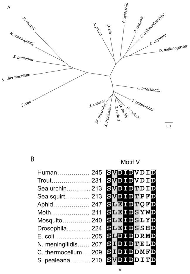
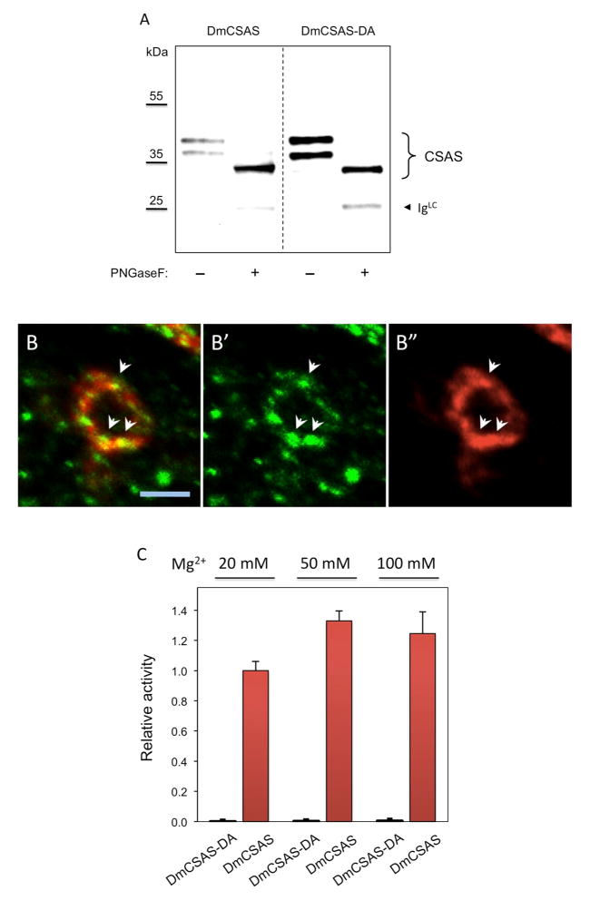
CMP-sialic acid synthetase (CSAS) is a key enzyme of the sialylation pathway. CSAS produces the activated sugar donor, CMP-sialic acid, which serves as a substrate for sialyltransferases to modify glycan termini with sialic acid. Unlike other animal CSASs that normally localize in the nucleus, Drosophila melanogaster CSAS (DmCSAS) localizes in the cell secretory compartment, predominantly in the Golgi, which suggests that this enzyme has properties distinct from those of its vertebrate counterparts. To test this hypothesis, we purified recombinant DmCSAS and characterized its activity in vitro Our experiments revealed several unique features of this enzyme. DmCSAS displays specificity for N-acetylneuraminic acid as a substrate, shows preference for lower pH and can function with a broad range of metal cofactors. When tested at a pH corresponding to the Golgi compartment, the enzyme showed significant activity with several metal cations, including Zn(2+), Fe(2+), Co(2+) and Mn(2+), whereas the activity with Mg(2+) was found to be low. Protein sequence analysis and site-specific mutagenesis identified an aspartic acid residue that is necessary for enzymatic activity and predicted to be involved in co-ordinating a metal cofactor. DmCSAS enzymatic activity was found to be essential in vivo for rescuing the phenotype of DmCSAS mutants. Finally, our experiments revealed a steep dependence of the enzymatic activity on temperature. Taken together, our results indicate that DmCSAS underwent evolutionary adaptation to pH and ionic environment different from that of counterpart synthetases in vertebrates. Our data also suggest that environmental temperatures can regulate Drosophila sialylation, thus modulating neural transmission.

Keywords: CMP-sialic acid synthetase; Drosophila glycosylation; enzyme evolution; sialic acid; sialylation.


Conflict of interest statement

The authors declare that they have no conflicts of interest with the contents of this article.

Figures

Figure 1
Figure 1
Purification of Drosophila CSAS protein and its in vitro enzymatic activity. A, DmCSAS-FLAG protein purified on FLAG-affinity beads and analyzed by Coomassie staining of SDS-PAGE gels and western blot. Lane 1, non-specific proteins purified from S2 cells without DmCSAS-FLAG expression. Lane 2, DmCSAS-FLAG purified from S2 cells. Lane 3: western blot analysis of purified DmCSAS (same sample as on lane 2). The positions of two DmCSAS glycoforms, as well as heavy and light chains of FLAG antibody leached from the beads are indicated. B, HPLC chromatograms illustrating the detection of CMP-Neu5Ac. The top trace shows CMP-Neu5Ac acid standard as a control. Traces below show the gradual increase of CMP-Neu5Ac product over a period of 90 min in the assay with purified DmCSAS. C, Purified DmCSAS can stably produce CMP-Neu5Ac for up to 90 min. The assays were carried our at standard reaction conditions (100mM Tris-HCl pH 8.0, 20mM MgCl2, 0.2 mM DTT, 3 mM sialic acid, 5.5 mM CTP, and 100 ng DmCSAS) at 40°C.
Figure 2
Figure 2
Effect of temperature and pH on DmCSAS activity. A, The dependence of enzyme activity on temperature. B, The dependence of enzyme activity on pH. Unless indicated otherwise, assays were performed at standard reaction conditions in the presence of 20 mM Mg2+ at 37°C, pH 8. Cacodylate buffer was used in assays with pH ≤ 7.4 (diamonds) and Tris-HCl buffer was used at pH ≥ 7.2 (squares).
Figure 3
Figure 3
Kinetic analysis of DmCSAS and comparison of substrate specificities of Drosophila and human CMP-Sia synthetases. AC, Graphs show kinetics for CTP (A), Neu5Ac (B), and Neu5Gc (C). D–E, Substrate specificities of purified DmCSAS (D) and HsCSAS (E) with Neu5Ac, Neu5Gc, KDN and KDO as tested substrates. Unless indicated otherwise, assays were performed using standard reaction conditions that included 100mM Tris-HCl pH 8.0, 20mM MgCl2, 0.2 mM DTT, 3 mM sugar substrate, and 5.5 mM CTP. Drosophila and human enzymes were used at 100 ng and 30 ng per reaction, respectively. Reactions were incubated for 1 hour at 37°C. The identity of CMP-Neu5Ac and CMP-Neu5Gc products was confirmed by MS analyses (see Supplementary Materal, Fig. S1 and Table S1).
Figure 4
Figure 4
Effect of metal ions on CMP-Sia synthetase activity. A, DmCSAS requires the presence of divalent metal cations for its activity. The enzyme was assayed at different concentrations of Mg2+. B, DmCSAS can utilize different metal cofactors, while metal cofactors have differential effects on DmCSAS activity at distinct pH. C, Human CMP-Sia synthetase assayed with different metal ions at the same reaction conditions as the Drosophila enzyme in B. DE, Substrate specificity (D) and temperature dependence (E) of DmCSAS activity in presence Mn2+ as a cofactor at pH 7.6. Unless indicated otherwise, assays were performed in standard reaction conditions using corresponding metal cofactor at 5 mM.
Figure 4
Figure 4
Effect of metal ions on CMP-Sia synthetase activity. A, DmCSAS requires the presence of divalent metal cations for its activity. The enzyme was assayed at different concentrations of Mg2+. B, DmCSAS can utilize different metal cofactors, while metal cofactors have differential effects on DmCSAS activity at distinct pH. C, Human CMP-Sia synthetase assayed with different metal ions at the same reaction conditions as the Drosophila enzyme in B. DE, Substrate specificity (D) and temperature dependence (E) of DmCSAS activity in presence Mn2+ as a cofactor at pH 7.6. Unless indicated otherwise, assays were performed in standard reaction conditions using corresponding metal cofactor at 5 mM.
Figure 5
Figure 5
Phylogenetic analysis and metal binding site comparison between CMP-Sia synthetases from different species. A, Phylogenetic tree illustrating evolutionary relationship between CMP-Sia synthetases from bacteria, insects and complex animals (deuterostomes). The tree was built by ClustalW2 program using neighbor-joining algorithm. The accession numbers of bacterial sequences: Pseudomonas veronii, WP_017848838.1; Shewanella pealeana, CP000851.1; Clostridium thermocellum, CP002416.1; Neisseria meningitidis, AAB60780.1; Escherichia coli, J05023.1. The accession numbers of insect proteins: Acyrthosiphon pisum (pea aphid), NP_001156112.1; Diaphorina citri (Asian citrus psyllid), XM_008472953.1; Plutella xylostella (moth), XM_011559466.1; Aedes aegypti (yellow fever mosquito), XM_001662967.1; Culex quinquefasciatus (southern house mosquito) XP_001842321.1; Ceratitis capitata (Mediterranean fruit fly), XM_004522975.1. The accession numbers of deuterostome proteins: Ciona intestinalis (sea squirt), NM_001100127.1; Strongylocentrotus purpuratus (sea urchin) CSAS NM_001126308.1; Danio rerio (zebrafish) JQ015186.1, JQ015187.1; Oncorhynchus mykiss (rainbow trout), NM_001124190.1; Xenopus tropicalis (frog), NM_001097281.1; Mus musculus (mouse), NP_034038.2; Homo sapiens (human), NP_061156.1. B, The fragment of CMP-Sia synthetase multiple sequence alignment showing the conserved Motif V involved in metal cofactor coordination. Asterisk indicates the aspartic acid residue of the DID triad that is conserved in bacterial and vertebrate sequences but is substituted by glutamic acid in insect CSAS proteins. The multiple alignments were generated by Clustal Omega server at EMBL.
Figure 6
Figure 6
Analyses of the DmCSAS-DA mutant protein. A, DmCSAS-DA (DmCSAS with D228->A substitution) is properly glycosylated, as indicated by PNGase F treatment. DmCSAS wildtype protein was used as a control. Proteins were purified on FLAG affinity beads, treated with PNGase F, and analyzed by SDS-PAGE followed by western blot detection. Both proteins are expressed as two glycoforms represented by two bands on the gel. Upon PNGaseF – mediated removal of N-glycans, these bands collapse into one band of a lower molecular mass corresponding to deglycosylated protein. B, DmCSAS-DA is localized in the Golgi compartment when expressed in the CNS neurons in vivo, as reveled by double immunofluorescent staining with the GM130 Golgi marker [47]. B′ and B″ show single channel staining for GM130 (green) and DmCSAS-DA (red), respectively. B is the overlay of B′ and B″. Arrows point at examples of co-localization between GM130 and DmCSAS-DA. DmCSAS-DA was expressed in the CNS using UAS-GAL4 system. Images of fixed, dissected and stained brains were obtained using epifluorescent microscopy with optical sectioning. The image shows a confocal section through the cell body of a single neuron. Scale bar is 5 μm. C, CMP-Sia synthetase activity assays of DmCSAS-DA mutant at increasing concentrations of Mg2+. DmCSAS wildtype protein was used as a positive control. No enzymatic activity of DmCSAS-DA was detected. D, DmCSAS activity was tested in vivo using a transgenic rescue approach. DmCSAS-DA and DmCSAS wildtype proteins were expressed in DmCSAS homozygous null mutants (DmCSAS) using UAS-GAL4 system. The rescue of DmCSAS phenotype was analyzed by TS-paralysis assays. At least 20 flies were assayed for each genotype. o and *** indicate not statistically significant and highly significant differences, respectively. Error bars represent SEM in all panels.
Figure 6
Figure 6
Analyses of the DmCSAS-DA mutant protein. A, DmCSAS-DA (DmCSAS with D228->A substitution) is properly glycosylated, as indicated by PNGase F treatment. DmCSAS wildtype protein was used as a control. Proteins were purified on FLAG affinity beads, treated with PNGase F, and analyzed by SDS-PAGE followed by western blot detection. Both proteins are expressed as two glycoforms represented by two bands on the gel. Upon PNGaseF – mediated removal of N-glycans, these bands collapse into one band of a lower molecular mass corresponding to deglycosylated protein. B, DmCSAS-DA is localized in the Golgi compartment when expressed in the CNS neurons in vivo, as reveled by double immunofluorescent staining with the GM130 Golgi marker [47]. B′ and B″ show single channel staining for GM130 (green) and DmCSAS-DA (red), respectively. B is the overlay of B′ and B″. Arrows point at examples of co-localization between GM130 and DmCSAS-DA. DmCSAS-DA was expressed in the CNS using UAS-GAL4 system. Images of fixed, dissected and stained brains were obtained using epifluorescent microscopy with optical sectioning. The image shows a confocal section through the cell body of a single neuron. Scale bar is 5 μm. C, CMP-Sia synthetase activity assays of DmCSAS-DA mutant at increasing concentrations of Mg2+. DmCSAS wildtype protein was used as a positive control. No enzymatic activity of DmCSAS-DA was detected. D, DmCSAS activity was tested in vivo using a transgenic rescue approach. DmCSAS-DA and DmCSAS wildtype proteins were expressed in DmCSAS homozygous null mutants (DmCSAS) using UAS-GAL4 system. The rescue of DmCSAS phenotype was analyzed by TS-paralysis assays. At least 20 flies were assayed for each genotype. o and *** indicate not statistically significant and highly significant differences, respectively. Error bars represent SEM in all panels.

Similar articles

Cited by

References

    1. Varki A, Schauer R. Sialic Acids. In: Varki A, Cummings RD, Esko JD, Freeze HH, Stanley P, Bertozzi CR, Hart GW, Etzler ME, editors. Essentials of Glycobiology. Cold Spring Harbor Laboratory Press; Cold Spring Harbor, NY: 2009. pp. 199–219. - PubMed
    1. Varki A. Sialic acids as ligands in recognition phenomena. FASEB J. 1997;11:248–255. - PubMed
    1. Varki A. Sialic acids in human health and disease. Trends Mol Med. 2008;14:351–360. - PMC - PubMed
    1. Sellmeier M, Weinhold B, Münster-Kühnel A. CMP-Sialic Acid Synthetase: The Point of Constriction in the Sialylation Pathway. Springer; Berlin Heidelberg: 2013. pp. 1–29. - PubMed
    1. Freeze HH, Elbein AD. Glycosylation Precursors. In: Varki A, Cummings RD, Esko JD, Freeze HH, Stanley P, Bertozzi CR, Hart GW, Etzler ME, editors. Essentials of Glycobiology. Cold Spring Harbor Laboratory Press; Cold Spring Harbor, NY: 2009. pp. 47–61.

Publication types

MeSH terms

LinkOut - more resources