Review
. 2016 Apr 26;8(4):136-57.
doi: 10.4252/wjsc.v8.i4.136.

Migration of bone marrow progenitor cells in the adult brain of rats and rabbits

Affiliations
Review

Migration of bone marrow progenitor cells in the adult brain of rats and rabbits

Donnahue Dennie et al. World J Stem Cells. .

Abstract

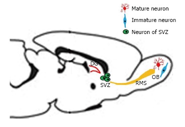
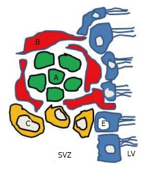
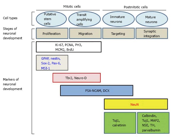
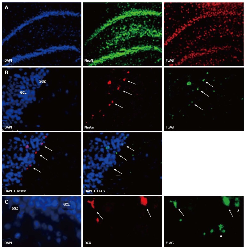
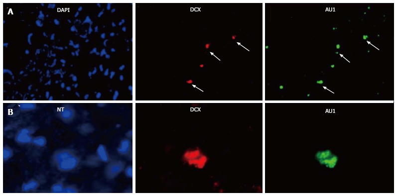
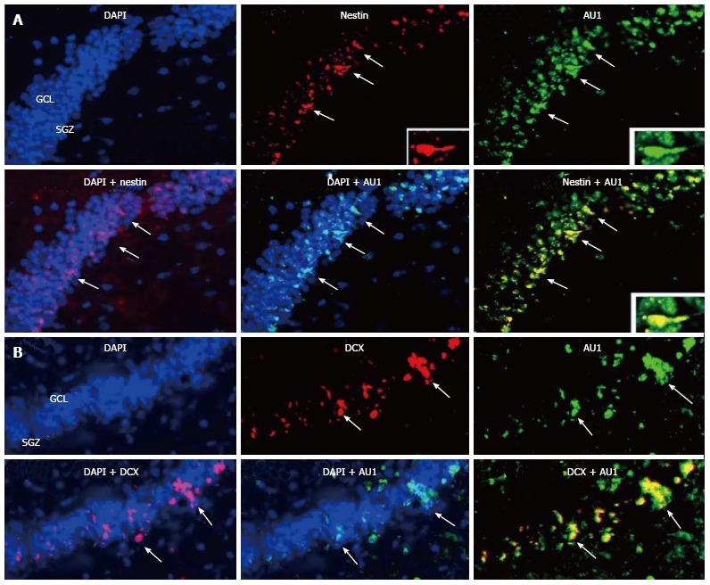
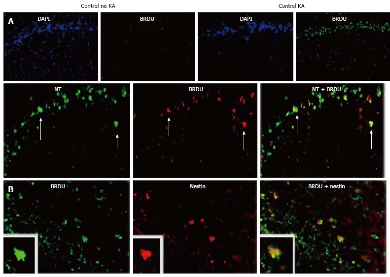
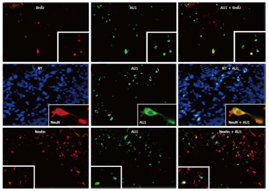
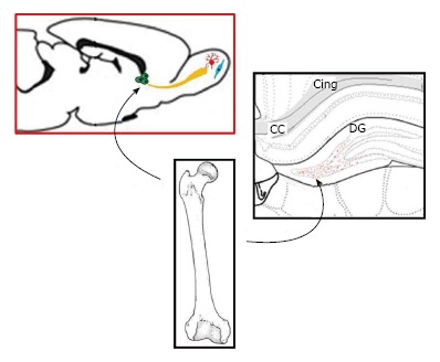
Neurogenesis takes place in the adult mammalian brain in three areas: Subgranular zone of the dentate gyrus (DG); subventricular zone of the lateral ventricle; olfactory bulb. Different molecular markers can be used to characterize the cells involved in adult neurogenesis. It has been recently suggested that a population of bone marrow (BM) progenitor cells may migrate to the brain and differentiate into neuronal lineage. To explore this hypothesis, we injected recombinant SV40-derived vectors into the BM and followed the potential migration of the transduced cells. Long-term BM-directed gene transfer using recombinant SV40-derived vectors leads to expression of the genes delivered to the BM firstly in circulating cells, then after several months in mature neurons and microglial cells, and thus without central nervous system (CNS) lesion. Most of transgene-expressing cells expressed NeuN, a marker of mature neurons. Thus, BM-derived cells may function as progenitors of CNS cells in adult animals. The mechanism by which the cells from the BM come to be neurons remains to be determined. Although the observed gradual increase in transgene-expressing neurons over 16 mo suggests that the pathway involved differentiation of BM-resident cells into neurons, cell fusion as the principal route cannot be totally ruled out. Additional studies using similar viral vectors showed that BM-derived progenitor cells migrating in the CNS express markers of neuronal precursors or immature neurons. Transgene-positive cells were found in the subgranular zone of the DG of the hippocampus 16 mo after intramarrow injection of the vector. In addition to cells expressing markers of mature neurons, transgene-positive cells were also positive for nestin and doublecortin, molecules expressed by developing neuronal cells. These cells were actively proliferating, as shown by short term BrdU incorporation studies. Inducing seizures by using kainic acid increased the number of BM progenitor cells transduced by SV40 vectors migrating to the hippocampus, and these cells were seen at earlier time points in the DG. We show that the cell membrane chemokine receptor, CCR5, and its ligands, enhance CNS inflammation and seizure activity in a model of neuronal excitotoxicity. SV40-based gene delivery of RNAi targeting CCR5 to the BM results in downregulating CCR5 in circulating cells, suggesting that CCR5 plays an important role in regulating traffic of BM-derived cells into the CNS, both in the basal state and in response to injury. Furthermore, reduction in CCR5 expression in circulating cells provides profound neuroprotection from excitotoxic neuronal injury, reduces neuroinflammation, and increases neuronal regeneration following this type of insult. These results suggest that BM-derived, transgene-expressing, cells can migrate to the brain and that they become neurons, at least in part, by differentiating into neuron precursors and subsequently developing into mature neurons.

Keywords: Bone marrow; Brain; Cell therapy; Development; Doublecortin; Epilepsy; Hippocampus; Nestin; Neurons; SV40; Seizures; Stem cells.


Figures

Figure 1
Figure 1
Different areas of neurogenesis in the adult rodent. The three areas of neurogenesis in the adult are the dentate gyrus of the hippocampus, the subventricular zone, and the olfactory bulb. Some progenitor cells are migrating from the SVZ to the OB, along a rostral migratory stream. DG: Dentate gyrus; SVZ: Subventricular zone; OB: Olfactory bulb; RMS: Rostral migratory stream.
Figure 2
Figure 2
Four different types of progenitor cells in the subventricular zone. Type-A cells are migrating towards the olfactory bulb along the rostral migratory stream. Type B cells are believed to be the neural precursor cells and will give rise to both type C and type A cells. Type C cells are rapidly dividing immature precursor cells arranged in clusters along the migrating chains, and they are found only in the SVZ. E cells belong to ependymocytes lineage. LV: Lateral ventricle; SVZ: Subventricular zone.
Figure 3
Figure 3
Different markers of neuronal development in mitotic and postmitotic cells (modified from Ref. [21,35]). PSA-NCAM: Polysialated form of the neural cell adhesion molecule; DCX: Doublecortin; GFAP: Glial fibrillary acidic protein; PCNA: Proliferating cell nuclear antigen; PH3: Phosphorylated form of histone 3; MCM2: Minichromosome maintenance protein 2; MSI-1: Musashi-1; MAP2: Microtubule-associated protein 2; NSE: Neuron specific enolase; TH: Tyrosine hydroxylase.
Figure 4
Figure 4
Bone marrow derived cells can migrate to the rat normal hippocampus. Sixteen months after injection of SV (Nef-FLAG) into the rat bone marrow (BM), transgene expressing cells were detected in the dentate gyrus (DG). A: FLAG-positive cells colocalized with NeuN, a marker of mature neurons. FLAG+/NeuN+ cells were located in the granular cell layer (GCL), as well as in the subgranular zone (SGZ) and the hilus. No FLAG-expressing cells were detected in the brain after injection of SV (BUGT), a control vector, in the BM; B: Nestin+/FLAG+ cells were detected mainly at the border SGZ/GCL, and more rarely in the GCL (arrows); C: DCX+ positive cells expressing FLAG were seen at the border SGZ/GCL and more rarely in the GCL (arrows). Note that some FLAG+ cells were DCX-, and were probably mature neurons (arrowheads). In all experiments, nuclei were stained in blue by Vectashield containing DAPI.
Figure 5
Figure 5
Transgene (AU1) expression in the hippocampus of rats whose bone marrow has been injected with SV (RevM10.AU1). A: Almost all DCX-positive cells were expressing AU1 (arrows); B: DCX+/AU1+ cells were of neuronal lineage, because they were stained by NT (arrows). No AU1 expression was seen after bone marrow injection of SV (BUGT), a control vector. DCX: Doublecortin; NT: Neurotrace.
Figure 6
Figure 6
Migration of bone marrow derived cells to the rabbit hippocampus. Sixteen months after injection of SV (RevM10.AU1) into the rabbit bone marrow (BM), transgene-positive cells were seen in the dentate gyrus. Numerous AU1-positive cells colocalized with NeuN, a marker of postmitotic neurons (not shown). A: Nestin+/AU1+ cells were seen mainly at the border SGZ/GCL, and more rarely in the GCL (arrows); B: DCX+ positive cells expressing AU1 were detected at the border SGZ/GCL and more rarely in the GCL (arrows). SGZ: Subgranular zone; GCL: Granular cell layer; DCX: Doublecortin.
Figure 7
Figure 7
Kainic acid-induced regeneration in the hippocampus. Rats were injected with kainic acid (KA) intraperitoneally (i.p.), 10 mg/kg, and their hippocampi (HCs) analyzed by immunomicroscopy 7 d thereafter. A: Neurogenesis following KA treatment. Rats given KA or saline were injected with BrdU. Then, at 7 d post-KA, their DGs were immunostained for BrdU to visualize proliferating cells. DAPI counterstain is shown to facilitate interpretation. Arrows show neurons positive for BrDU. Double staining with Neurotrace (NT) for neurons is shown below; B: Double staining for nestin, a marker of proliferating and migrating neural cells (modified from Ref. [132]).
Figure 8
Figure 8
RNAiR5 gene delivery to the bone marrow increases numbers of bone marrow-derived cells in the hippocampus and neuroproliferative activity in the hippocampus, in the resting state and in response to kainic acid treatment. Bone marrow-derived cells in the hippocampus (HC) were all neurons, and most proliferating cells in the HC were bone marrow-derived. Double immunostaining for AU1 plus BrdU (upper row) and nestin (lower row), and neuron identification using Neurotrace (NT) and NeuN (middle row) was performed. Shown are HC sections from kainic acid-treated rats, transduced with SV (RNAiR5-RevM10.AU1). Most of the AU1+ cells were also positive for BrdU (79.8%, ± 6.9%) as a marker of cell proliferation, and most BrdU+ cells were also AU1+ (72.2%, ± 7.4%). Numerous AU1+ cells were also positive for nestin, a marker of proliferating and migrating neurons, although there were as well many nestin-positive cells that did not express AU1. The equivalence of NT identification of neurons and NeuN immunostaining of neurons is illustrated in the insets in the middle row. Data are representative of 3 independent experiments (modified from Ref. [132]).
Figure 9
Figure 9
Figure suggesting the participation of bone marrow progenitor cells to neurogenesis in adults. DG: Dentate gyrus.

References

    1. Nelson DM, Metzger ME, Donahue RE, Morgan RA. In vivo retrovirus-mediated gene transfer into multiple hematopoietic lineages in rabbits without preconditioning. Hum Gene Ther. 1997;8:747–754. - PubMed
    1. Porada CD, Tran ND, Zhao Y, Anderson WF, Zanjani ED. Neonatal gene therapy. transfer and expression of exogenous genes in neonatal sheep following direct injection of retroviral vectors into the bone marrow space. Exp Hematol. 2000;28:642–650. - PubMed
    1. McCauslin CS, Wine J, Cheng L, Klarmann KD, Candotti F, Clausen PA, Spence SE, Keller JR. In vivo retroviral gene transfer by direct intrafemoral injection results in correction of the SCID phenotype in Jak3 knock-out animals. Blood. 2003;102:843–848. - PubMed
    1. Mátrai J, Chuah MK, VandenDriessche T. Recent advances in lentiviral vector development and applications. Mol Ther. 2010;18:477–490. - PMC - PubMed
    1. Frecha C, Costa C, Nègre D, Amirache F, Trono D, Rio P, Bueren J, Cosset FL, Verhoeyen E. A novel lentiviral vector targets gene transfer into human hematopoietic stem cells in marrow from patients with bone marrow failure syndrome and in vivo in humanized mice. Blood. 2012;119:1139–1150. - PubMed

LinkOut - more resources