

Relationship between Spinal Hemangioblastoma Location and Age
- PMID: 27114772
- PMCID: PMC4843068
- DOI: 10.4184/asj.2016.10.2.309
Relationship between Spinal Hemangioblastoma Location and Age
Abstract
Study design: Retrospective case series.
Purpose: To investigate the relationship between tumor location and clinical characteristics.
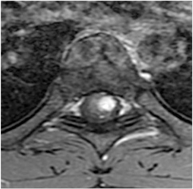
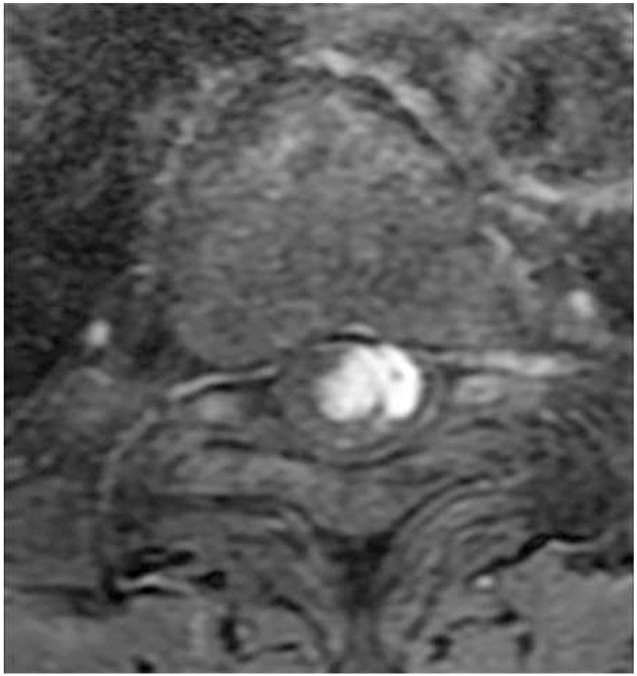
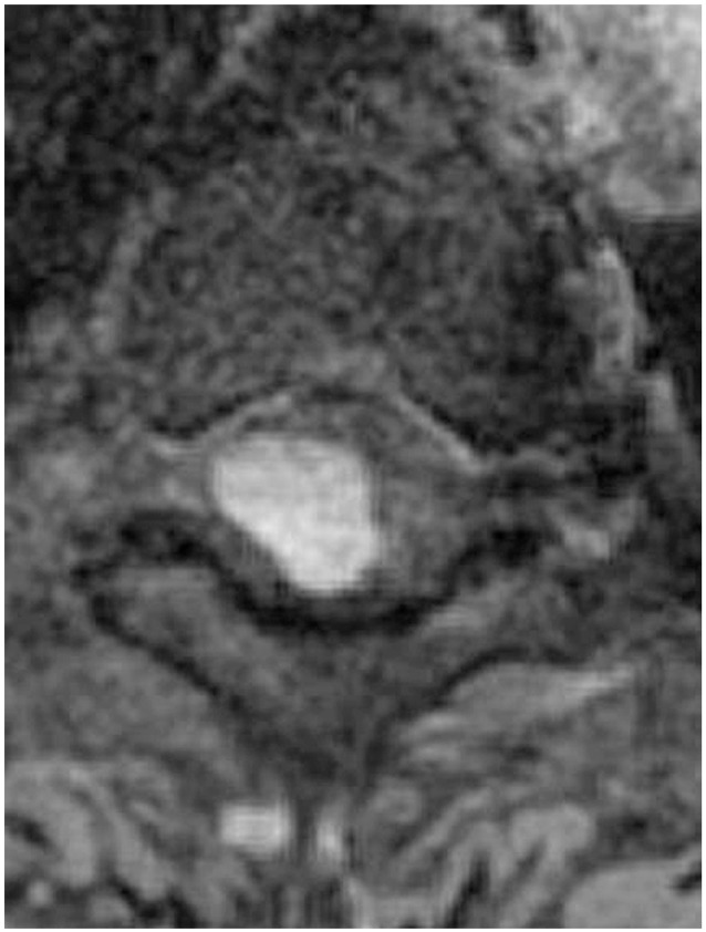
Overview of literature: Hemangioblastoma is a rare disease that develops in the central nervous system. Magnetic resonance imaging (MRI) is useful to evaluate hemangioblastomas. Hemangioblastoma's location is designated as intramedullary, intramedullary+extramedullary, or extramedullary by MRI.
Methods: We analyzed 11 patients who underwent surgery for spinal hemangioblastoma. Using T1 contrast axial MRI data, the cases were divided into three groups (intramedullary, intramedullary+extramedullary, and extramedullary). Patient demographics, MRI findings, and preoperative neurological status were analyzed and compared for each group.
Results: The average age of patients with intramedullary, intramedullary+extramedullary, and extramedullary hemangioblastoma was 34.0, 64.4, and 67.5 years, respectively. Patients in the intramedullary hemangioblastoma group were younger than the other groups. Extramedullary cases had a smaller syrinx compared to the other groups.
Conclusions: Age may play an important role in the hemangioblastoma tumor location and the subsequent diagnosis by an MRI.
Keywords: Age; Magnetic resonance imaging; Spinal hemangioblastoma; Tumor location.
Conflict of interest statement
Figures
Similar articles
-
Differentiation of localization of spinal hemangioblastomas based on imaging and pathological findings.Eur Spine J. 2011 Aug;20(8):1377-84. doi: 10.1007/s00586-011-1814-6. Epub 2011 Apr 29. Eur Spine J. 2011. PMID: 21528401 Free PMC article.
-
Rapid Worsening of Symptoms and High Cell Proliferative Activity in Intra- and Extramedullary Spinal Hemangioblastoma: A Need for Earlier Surgery.Global Spine J. 2017 Feb;7(1):6-13. doi: 10.1055/s-0036-1580612. Epub 2017 Feb 1. Global Spine J. 2017. PMID: 28451503 Free PMC article.
-
Prognostic Factors of Spinal Intramedullary Hemangioblastoma : Analysis of Surgical Outcomes and Tumor Characteristics.J Korean Neurosurg Soc. 2024 Nov;67(6):637-645. doi: 10.3340/jkns.2023.0221. Epub 2024 Apr 23. J Korean Neurosurg Soc. 2024. PMID: 38650430 Free PMC article.
-
Sporadic Intradural Extramedullary Hemangioblastoma of the Cauda Equina: Case Report and Literature Review.World Neurosurg. 2018 Jan;109:436-441. doi: 10.1016/j.wneu.2017.10.104. Epub 2017 Oct 28. World Neurosurg. 2018. PMID: 29107720 Review.
-
Spinal cord hemangioblastoma with extensive syringomyelia.J Chin Med Assoc. 2005 Jan;68(1):40-4. doi: 10.1016/S1726-4901(09)70131-5. J Chin Med Assoc. 2005. PMID: 15742863 Review.
Cited by
-
Hydrocephalus: a rare initial manifestation of sporadic intramedullary hemangioblastoma : Intramedullary hemangioblastoma presenting as hydrocephalus.Childs Nerv Syst. 2017 Aug;33(8):1399-1403. doi: 10.1007/s00381-017-3415-0. Epub 2017 Apr 25. Childs Nerv Syst. 2017. PMID: 28444460
-
Neurological Outcome of Spinal Hemangioblastomas: An International Observational Multicenter Study About 35 Surgical Cases.Cancers (Basel). 2025 Apr 24;17(9):1428. doi: 10.3390/cancers17091428. Cancers (Basel). 2025. PMID: 40361357 Free PMC article.
-
Intradural extramedullary hemangioblastoma of the thoracic cord: A case report.Surg Neurol Int. 2021 Mar 30;12:126. doi: 10.25259/SNI_795_2020. eCollection 2021. Surg Neurol Int. 2021. PMID: 33880231 Free PMC article.
-
Intradural Extramedullary Hemangioblastoma of the Cervical Spine: Case Report and Literature Review.Cureus. 2022 May 18;14(5):e25125. doi: 10.7759/cureus.25125. eCollection 2022 May. Cureus. 2022. PMID: 35733499 Free PMC article.
-
Immunotherapy for Pediatric Brain and Spine Tumors: Current State and Future Directions.Pediatr Neurosurg. 2023;58(5):313-336. doi: 10.1159/000528792. Epub 2022 Dec 22. Pediatr Neurosurg. 2023. PMID: 36549282 Free PMC article. Review.
References
-
- Lee DK, Choe WJ, Chung CK, Kim HJ. Spinal cord hemangioblastoma: surgical strategy and clinical outcome. J Neurooncol. 2003;61:27–34. - PubMed
-
- Mandigo CE, Ogden AT, Angevine PD, McCormick PC. Operative management of spinal hemangioblastoma. Neurosurgery. 2009;65:1166–1177. - PubMed
-
- Glasker S, Berlis A, Pagenstecher A, Vougioukas VI, Van Velthoven V. Characterization of hemangioblastomas of spinal nerves. Neurosurgery. 2005;56:503–509. - PubMed
-
- Kanno H, Yamamoto I, Nishikawa R, et al. Spinal cord hemangioblastomas in von Hippel-Lindau disease. Spinal Cord. 2009;47:447–452. - PubMed
LinkOut - more resources
Full Text Sources
Other Literature Sources
Research Materials
