. 2016 Nov 1;194(9):1140-1151.
doi: 10.1164/rccm.201510-1955OC.

A Selective Transforming Growth Factor-β Ligand Trap Attenuates Pulmonary Hypertension

Affiliations

A Selective Transforming Growth Factor-β Ligand Trap Attenuates Pulmonary Hypertension

Lai-Ming Yung et al. Am J Respir Crit Care Med. .

Abstract

Rationale: Transforming growth factor-β (TGF-β) ligands signal via type I and type II serine-threonine kinase receptors to regulate broad transcriptional programs. Excessive TGF-β-mediated signaling is implicated in the pathogenesis of pulmonary arterial hypertension, based in part on the ability of broad inhibition of activin-like kinase (ALK) receptors 4/5/7 recognizing TGF-β, activin, growth and differentiation factor, and nodal ligands to attenuate experimental pulmonary hypertension (PH). These broad inhibition strategies do not delineate the specific contribution of TGF-β versus a multitude of other ligands, and their translation is limited by cardiovascular and systemic toxicity.

Objectives: We tested the impact of a soluble TGF-β type II receptor extracellular domain expressed as an immunoglobulin-Fc fusion protein (TGFBRII-Fc), serving as a selective TGF-β1/3 ligand trap, in several experimental PH models.

Methods: Signaling studies used cultured human pulmonary artery smooth muscle cells. PH was studied in monocrotaline-treated Sprague-Dawley rats, SU5416/hypoxia-treated Sprague-Dawley rats, and SU5416/hypoxia-treated C57BL/6 mice. PH, cardiac function, vascular remodeling, and valve structure were assessed by ultrasound, invasive hemodynamic measurements, and histomorphometry.

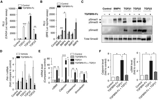
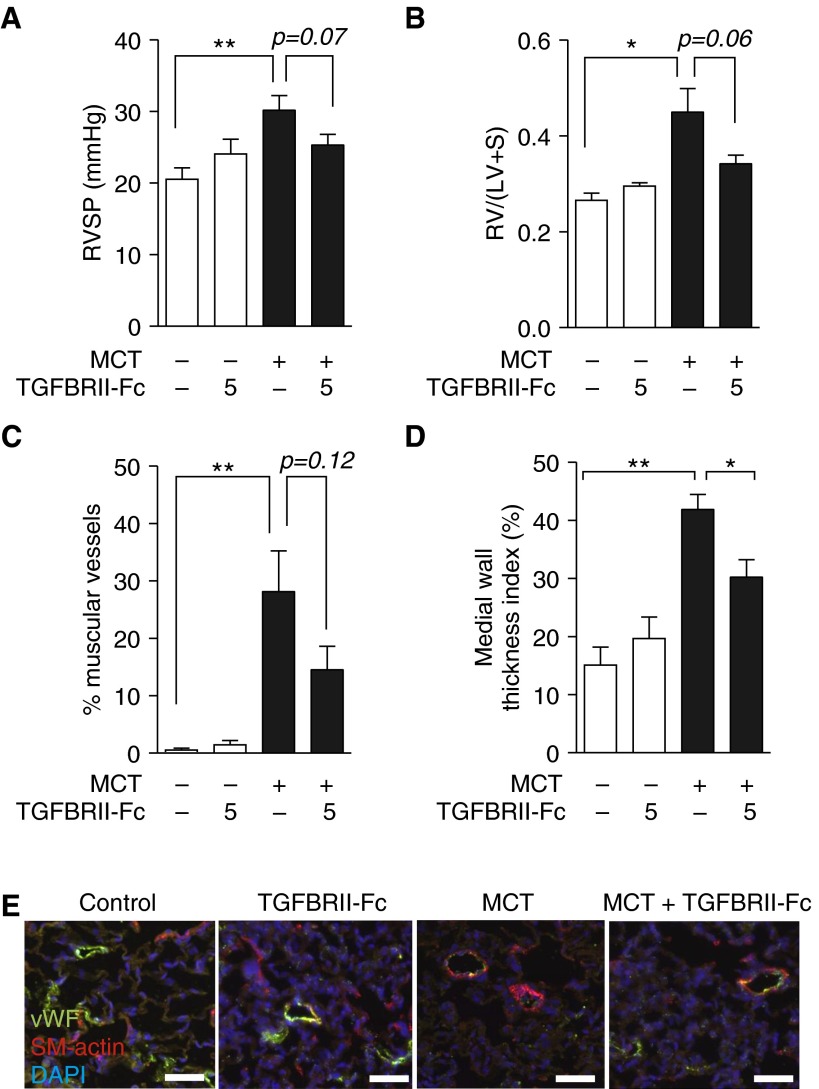
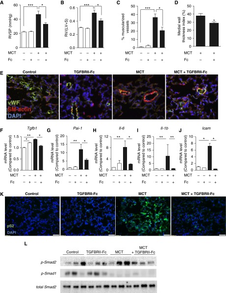
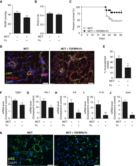
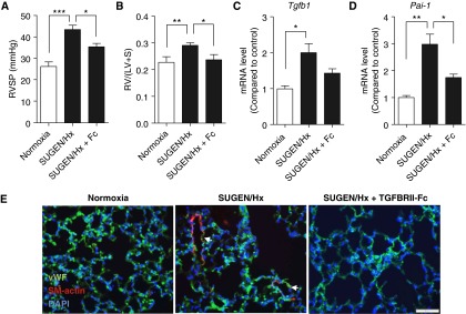
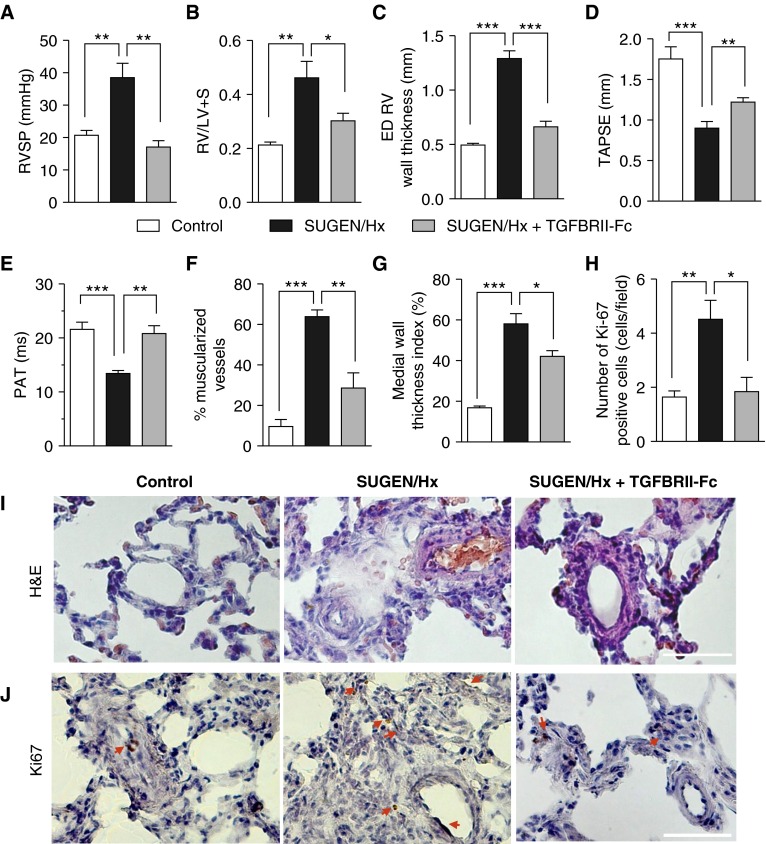
Measurements and main results: TGFBRII-Fc is an inhibitor of TGF-β1 and TGF-β3, but not TGF-β2, signaling. In vivo treatment with TGFBRII-Fc attenuated Smad2 phosphorylation, normalized expression of plasminogen activator inhibitor-1, and mitigated PH and pulmonary vascular remodeling in monocrotaline-treated rats, SU5416/hypoxia-treated rats, and SU5416/hypoxia-treated mice. Administration of TGFBRII-Fc to monocrotaline-treated or SU5416/hypoxia-treated rats with established PH improved right ventricular systolic pressures, right ventricular function, and survival. No cardiac structural or valvular abnormalities were observed after treatment with TGFBRII-Fc.

Conclusions: Our findings are consistent with a pathogenetic role of TGF-β1/3, demonstrating the efficacy and tolerability of selective TGF-β ligand blockade for improving hemodynamics, remodeling, and survival in multiple experimental PH models.

Keywords: pulmonary artery; pulmonary hypertension; transforming growth factor-β; vascular remodeling; vascular smooth muscle cells.


Figures

Figure 1.
Figure 1.
TGFBRII-Fc selectively inhibits transforming growth factor-β1 (TGF-β1)- and TGF-β3–induced signaling and blocks TGF-β–induced phenotypic modulation of smooth muscle cells. (A) HEK293 cells stably expressing a TGF-β–responsive CAGA-luciferase (Luc) reporter transgene and (B) C2C12 cells stably expressing a bone morphogenetic protein (BMP) response element reporter (BRE)–Luc reporter transgene were incubated in the presence or absence of TGFBRII-Fc (2 μg/ml) for 30 minutes before stimulation with various BMP ligands (10 ng/ml) or TGF-β1, -2, or -3 (1 ng/ml) overnight, revealing selective inhibition by TGFBRII-Fc of TGF-β1– and TGF-β3– but not TGF-β2–induced activation of CAGA-luciferase activity, and slightly increased BRE-luciferase activity in response to BMP2 and BMP4 in the presence of TGFBRII-Fc. Human pulmonary artery smooth muscle cells were deprived of serum overnight, pretreated with TGFBRII-Fc (2 μg/ml), followed by incubation with BMP4 (10 ng/ml), TGF-β1, -2, or -3 (1 ng/ml of each for 30 min), and analyzed by immunoblot for (C) phosphorylated Smads (pSmad) 1, 2, and 3 and (D) for mRNA expression of the TGF-β transcriptional target PAI-1 by quantitative reverse transcriptase polymerase chain reaction. TGFBRII-Fc inhibited SMAD2 and SMAD3 activation, CAGA-Luc reporter activity, and Pai-1 mRNA expression via TGF-β1 and TGF-β3, but not TGF-β2. After stimulation with TGF-β1, the relative levels of (E) mRNA and (F) protein expression of smooth muscle cell contractile markers were examined at 24 and 72 hours, respectively. Data are expressed as mean ± SEM. *P < 0.05; **P < 0.01; ***P < 0.001 as indicated. αSMA = α-smooth muscle actin; PAI-1 = plasminogen activator inhibitor 1; RLU = relative light units.
Figure 2.
Figure 2.
Lower-dose treatment with TGFBRII-Fc elicits trends toward improved pulmonary hypertension, right ventricular hypertension (RVH), and pulmonary vascular remodeling. (A) Three weeks after treatment with monocrotaline (MCT) with or without TGFBRII-Fc (5 mg/kg, twice weekly i.p.), rats were analyzed in a blinded fashion to determine right ventricular systolic pressure (RVSP). (B) The degree of RVH was assessed based on measurement of Fulton’s ratio [RV/(LV + S)]. (C) Muscularization of distal intraacinar vessels (10–50 μm diameter) was quantified, and the percentages of fully (circumferentially) muscularized vessels were calculated. (D) Medial wall thickness was calculated for all fully muscularized intraacinar vessels (10–50 μm diameter). Wall thickness index was calculated as index = (external diameter – internal diameter)/external diameter × 100. TGFBRII-Fc treatment under this dosing regimen trended toward decreased frequency of muscularized vessels, and significantly reduced medial wall thickness, based on 100 to 150 vessels per treatment group from 6 to 8 rats each. *P < 0.05 or **P < 0.01 versus control animals or otherwise as shown. (E) Immunofluorescence of lung sections for von Willebrand’s factor (vWF) to mark vessels and smooth muscle actin (SM-actin) revealed qualitatively increased muscularization of small (<50 μm) arterioles following MCT treatment, which was reduced by the administration of TGFBRII-Fc (scale bars = 50 μm). Nuclei were counterstained with 4′,6-diamidino-2-phenylindole (DAPI). Data are shown as mean ± SEM (n  =  6–8). *P < 0.05 and **P < 0.01 as indicated. LV = left ventricle; RV = right ventricle; S = septum.
Figure 3.
Figure 3.
Higher-dose treatment with TGFBRII-Fc prevented pulmonary hypertension and vascular remodeling. Adult rats treated with monocrotaline (MCT) (40 mg/kg s.c. × 1) were administered TGFBRII-Fc (15 mg/kg i.p. twice weekly, starting 1 day after MCT injection) or vehicle for 3 weeks. Treatment with TGFBRII-Fc significantly attenuated (A) right ventricular systolic pressure (RVSP) and (B) right ventricular hypertension in comparison to vehicle (n = 6–8). (C and D) TGBRII-Fc decreased the percentage of fully muscularized vessels (10–50 μm diameter) (C) and medial wall thickness, calculated as (external diameter – internal diameter)/external diameter × 100 (D), based on 89 to 127 vessels per treatment group from 6 to 8 rats each. (E) TGFBRII-Fc treatment reduced muscularization evident by smooth muscle actin staining of von Willebrand (vWF)+ small vessels. (F–J) TGFBRII-Fc treatment normalized the expression of Tgfb1(F), Pai-1 (G), Il-6 (H), Il1b (I), and Icam (J) in the lung tissues of MCT-treated rats (n = 3–5). (K) Immunohistochemistry of lung sections and (L) immunoblotting of whole-lung lysates demonstrated qualitatively increased p-Smad2 (pS2) expression in the lungs of MCT-treated animals, which was normalized by treatment with TGFBRII-Fc. Conversely, treatment with MCT robustly decreased expression of p-Smad1/5/8, which was not significantly affected by treatment with TGFBRII-Fc. Nuclei were counterstained with 4′,6-diamidino-2-phenylindole (DAPI). Data are expressed as mean ± SEM. *P < 0.05; **P < 0.01; ***P < 0.001 as indicated. Scale bars = 50 μm. Icam = intercellular adhesion molecule; i.p. = intraperitoneal; LV = left ventricle; Pai-1 = plasminogen activator inhibitor 1; RV = right ventricle; S = septum; s.c. = subcutaneous; Tgfb1 = transforming growth factor-β1.
Figure 4.
Figure 4.
Treatment with TGFBRII-Fc after establishment of pulmonary hypertension (PH) is associated with partial rescue of PH and mortality. After monocrotaline (MCT) treatment, rats were administered TGFBRII-Fc (15 mg/kg three times weekly) in a delayed fashion starting on Day 17, after the establishment of PH. Among surviving animals at 35 days, there was (A) significantly decreased right ventricular systolic pressure (RVSP) with TGFBRII-Fc treatment, but (B) no significant difference in right ventricular hypertension. Values are shown as mean ± SEM (n = 8–11 per group). (C) Kaplan-Meier analysis revealed improved survival in the TGFBRII-Fc–treated group (n = 18 per group). (D and E) TGFBRII-Fc treatment attenuated pulmonary vascular remodeling in rats with established PH. (F–J) Delayed treatment with TGFBRII-Fc reduced the expression of Tgfb1 (F), Pai-1 (G), Il6 (H), Il1b (I), and Icam (J) in the lung tissues of MCT-treated rats (n = 3–5). (K) TGFBRII-Fc treatment reduced elevated phospho-Smad2 (pS2) levels in MCT lungs. Nuclei were counterstained with 4′,6-diamidino-2-phenylindole (DAPI). Data are shown as mean ± SEM. *P < 0.05; **P < 0.01 as indicated. Scale bars = 50 μm. Icam = intercellular adhesion molecule; LV = left ventricle; Pai-1 = plasminogen activator inhibitor 1; RV = right ventricle; S = septum; SM-actin = smooth muscle actin; Tgfb1 = transforming growth factor-β1; vWF = von Willebrand factor.
Figure 5.
Figure 5.
Efficacy of TGFBRII-Fc in a murine model of pulmonary hypertension. Adult male mice were treated with SU5416 (SUGEN) and exposed to hypoxia for 3 weeks. (A and B) TGFBRII-Fc treatment (15 mg/kg, three times per week) reduced right ventricular systolic pressure (RVSP) (A) and prevented right ventricular hypertrophy (B) compared with vehicle-treated mice. (C and D) TGFBRII-Fc treatment trended toward reduced Tgfb1 (C) and prevented the up-regulation of Pai-1 (D) mRNA levels in lungs of SU5416/hypoxia–treated mice. (E) TGFBRII-Fc treatment ameliorated pulmonary vascular remodeling. Data are expressed as mean ± SEM (n = 6–8). *P < 0.05; **P < 0.01; ***P <  0.001 as indicated. Scale bars = 50 μm. DAPI = 4′,6-diamidino-2-phenylindole; Hx = hypoxia; LV = left ventricle; Pai-1 = plasminogen activator inhibitor 1; RV = right ventricle; S = septum; SM-actin = smooth muscle actin; Tgfb1 = transforming growth factor-β1; vWF = von Willebrand factor.
Figure 6.
Figure 6.
Treatment with TGFBRII-Fc reverses established pulmonary hypertension in SU5416 (SUGEN)/hypoxia–treated rats. Male adult Sprague-Dawley rats received a single subcutaneous injection of SU5416 and were subjected to normobaric hypoxia (FiO2 = 0.10) for 3 weeks, followed by maintenance in normoxia for 3 weeks, during which rats were randomized to receive either vehicle or TGFBRII-Fc (15 mg/kg, three times per week). (A–C) TGFBRII-Fc treatment reduced right ventricular systolic pressure (RVSP) (A), attenuated right ventricular hypertension (B), and reduced echocardiographic end-diastolic (ED) right ventricular free wall thickness (C) compared with vehicle-treated rats. (D and E) TGFBRII-Fc treatment improved echocardiographic measures of right ventricular function via tricuspid annular plane systolic excursion (TAPSE) (D) and of pulmonary hypertension via pulmonary acceleration time (PAT) (E) in SU5416/hypoxia–treated rats. There were no differences in pulmonary ejection time among various experimental groups (data not shown). (F, G, and I) TGBRII-Fc decreased the percentage of fully muscularized vessels (10–50 μm diameter) and medial wall thickness, calculated as (external diameter – internal diameter)/external diameter × 100, based on 50 to 85 vessels per treatment group from 5 to 6 rats each. (H and J) TGFBRII-Fc also reduced the frequency of Ki67-positive cells in the intima and adventitia (indicated by red arrows) seen in SU5416/hypoxia–treated rats. Data are expressed as mean ± SEM (n = 5–6). *P < 0.05; **P < 0.01; ***P < 0.001 as indicated. Scale bars = 50 μm. H&E = hematoxylin and eosin; Hx = hypoxia; LV = left ventricle; RV = right ventricle; S = septum.

Comment in

References

    1. Megalou AJ, Glava C, Oikonomidis DL, Vilaeti A, Agelaki MG, Baltogiannis GG, Papalois A, Vlahos AP, Kolettis TM. Transforming growth factor-β inhibition attenuates pulmonary arterial hypertension in rats. Int J Clin Exp Med. 2010;3:332–340. - PMC - PubMed
    1. Strelau J, Schober A, Sullivan A, Schilling L, Unsicker K. Growth/differentiation factor-15 (GDF-15), a novel member of the TGF-beta superfamily, promotes survival of lesioned mesencephalic dopaminergic neurons in vitro and in vivo and is induced in neurons following cortical lesioning. J Neural Transm Suppl. 2003;(65):197–203. - PubMed
    1. Deng Z, Morse JH, Slager SL, Cuervo N, Moore KJ, Venetos G, Kalachikov S, Cayanis E, Fischer SG, Barst RJ, et al. Familial primary pulmonary hypertension (gene PPH1) is caused by mutations in the bone morphogenetic protein receptor-II gene. Am J Hum Genet. 2000;67:737–744. - PMC - PubMed
    1. Lane KB, Machado RD, Pauciulo MW, Thomson JR, Phillips JA, III, Loyd JE, Nichols WC, Trembath RC International PPH Consortium. Heterozygous germline mutations in BMPR2, encoding a TGF-beta receptor, cause familial primary pulmonary hypertension. Nat Genet. 2000;26:81–84. - PubMed
    1. Waite KA, Eng C. From developmental disorder to heritable cancer: it’s all in the BMP/TGF-beta family. Nat Rev Genet. 2003;4:763–773. - PubMed

MeSH terms

Substances