Review
. 2016 Oct;1863(10):2503-14.
doi: 10.1016/j.bbamcr.2016.04.020. Epub 2016 Apr 23.

VDAC2-specific cellular functions and the underlying structure

Affiliations
Review

VDAC2-specific cellular functions and the underlying structure

Shamim Naghdi et al. Biochim Biophys Acta. 2016 Oct.

Abstract

Voltage Dependent Anion-selective Channel 2 (VDAC2) contributes to oxidative metabolism by sharing a role in solute transport across the outer mitochondrial membrane (OMM) with other isoforms of the VDAC family, VDAC1 and VDAC3. Recent studies revealed that VDAC2 also has a distinctive role in mediating sarcoplasmic reticulum to mitochondria local Ca(2+) transport at least in cardiomyocytes, which is unlikely to be explained simply by the expression level of VDAC2. Furthermore, a strictly isoform-dependent VDAC2 function was revealed in the mitochondrial import and OMM-permeabilizing function of pro-apoptotic Bcl-2 family proteins, primarily Bak in many cell types. In addition, emerging evidence indicates a variety of other isoform-specific engagements for VDAC2. Since VDAC isoforms display 75% sequence similarity, the distinctive structure underlying VDAC2-specific functions is an intriguing problem. In this paper we summarize studies of VDAC2 structure and functions, which suggest a fundamental and exclusive role for VDAC2 in health and disease. This article is part of a Special Issue entitled: Mitochondrial Channels edited by Pierre Sonveaux, Pierre Maechler and Jean-Claude Martinou.

Keywords: Apoptosis; Bak; Bax; Bid; Calcium; Mitochondria; Sarcoplasmic reticulum; VDAC2.


Figures

Fig. 1
Fig. 1
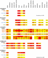
Protein sequence of different VDAC isoforms. A) Protein sequence comparison using Clustal Omega [144] among different VDAC isoforms including VDAC1 (NP_003365.1), VDAC2 (1): VDAC2 isoform1 (NP_001171712.1), VDAC2 (2): VDAC2 isoform2 (NP_003366.2), VDAC3 (1): VDAC3 isoform1 (NP_005653.3). VDAC3 isoform 2 (NP_001129166.1, not shown) is similar to VDAC3 isoform 1 with an extra methionine in the position of amino acid 40 (see arrow). B) Protein sequence comparison of VDAC2 in different species including human (Homo sapiens): NP_003366.2, monkey (Macaca mulatta): NP_ 001244353.1, cow (Bos taurus): NP_776911.2, pig (Sus scrofa): NP_999534.1, rat (Rattus norvegicous): NP_112644.1, rabbit (Oryctolagus cuniculus): NP_001076187.1, mouse (Mus musculus): NP_035825.1, frog (Xenopus laevis): NP_001089399.1, fish (Danio rerio): NP_955879.1, and chicken (Gallus gallus): NP_990072.1. NP code protein: Protein sequences that have been curated from Refseq database (an open access database from NCBI: national center for biotechnology information). The sequence has been colored by conservation using Jalview software and 33 was the assigned number for threshold.
Fig. 2
Fig. 2
High throughput data for hVDAC expression pattern in various tissues using different methods. A) RNA quantification from the brain, heart, liver and kidney using northern blot. The values are amol·μg−1/total RNA. B) Aceview: RNA-seq based gene expression profile in different tissues. Values are sFPKM, which stands for significant fragment per kilobase in one million read. C) BioGPS: tissue specific pattern of mRNA expression using customized Affymetrix high-density oligonucleotide biochip (dataset: GeneAtlas U133A). The probes used are 211662_s_at for VDAC2, 212038_s_at (a), 217139_at (b), 217140_s_at (c) for VDAC1 and 208845_ at (d), 208844_at (e), 208846_s_at (f) for VDAC3. The expression values are based on the fluorescence intensity and have been summarized using gcrma algorithms (https://www.bioconductor.org). Since each probe has different background characteristics, comparing the values from different probe sets is not recommended. In addition, it needs to be considered that one single probe might result in no similar result from study-to-study (comparing presented BioGPS data with information from coxpresdb database. (http://coxpresdb.jp). D) MitoCarta: Relative abundance of proteins reported in the MitoCarta Human database from different tissues. The values were determined by MS total peak intensity and can only be used to compare between tissues for a given protein. E) Western blot quantification from an antibody that recognizes all VDAC isoforms similarly. Therefore the levels of the different isoforms are comparable with each other. Values are expressed as the percentage of total VDACs in the mitochondria isolated from each tissue. Color intensity is set to darker color as higher number.
Fig. 3
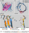
Fig. 3. VDAC2 specific 3D structure potentially relevant for Bak recruitment
A,B) Side and top view of zfV2 (PDB code: 4bum) illustrated using Pymol. Pink is the sufficient segment and yellow (sticks) is the necessary residues including S156 and D158. C) Upper Right: Side view of the segment containing amino acids S156 and D158 (yellow sticks) in zfV2 versus the relevant residues in mV1 (N156 and E158 orange sticks). Residues 111-167 in zfV2 (blue) and mV1 (PDB code: 3emn, orange) were superimposed. Upper Left: Interior view of the channels showing the residues,which are within 4Å distance from either S156 or D158 in zfV2 (sticks with carboncolored blue) or N156 or E158 in V1 (sticks with carbon colored orange). In sticks, oxygen is red and nitrogen is green. Lower panel: The adjacent residues have also been marked in the sequences of zfV2 (blue) and mV1 (orange). Reproduced with from Ref , by permission of PNAS.
Fig. 4
Fig. 4
Scheme of VDAC2 various functions/interaction partners. mtCU: Mitochondrial Calcium Uniporter, PTP: permeability transition pore, mtx: Metaxin.

References

    1. Dolder M, Zeth K, Tittmann P, Gross H, Welte W, Wallimann T. Crystallization of the human, mitochondrial voltage-dependent anion-selective channel in the presence of phospholipids. J. Struct. Biol. 1999;127:64–71. - PubMed
    1. Guo XW, Mannella CA. Conformational change in the mitochondrial channel, VDAC, detected by electron cryo-microscopy. Biophys. J. 1993;64:545–549. - PMC - PubMed
    1. Mannella CA, Kinnally KW. Reflections on VDAC as a voltage-gated channel and a mitochondrial regulator. J. Bioenerg. Biomembr. 2008;40:149–155. - PubMed
    1. Hiller S, Garces RG, Malia TJ, Orekhov VY, Colombini M, Wagner G. Solution structure of the integral human membrane protein VDAC-1 in detergent micelles. Science. 2008;321:1206–1210. - PMC - PubMed
    1. Colombini M. Pore size and properties of channels from mitochondria isolated from Neurospora crassa. J. Membr. Biol. 1980;53:79–84.

Publication types

MeSH terms

Substances

LinkOut - more resources