

Semi-supervised clustering of fractionated electrograms for electroanatomical atrial mapping
- PMID: 27117088
- PMCID: PMC4845510
- DOI: 10.1186/s12938-016-0154-5
Semi-supervised clustering of fractionated electrograms for electroanatomical atrial mapping
Abstract
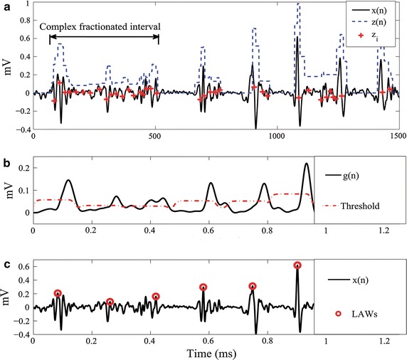
Background: Electrogram-guided ablation procedures have been proposed as an alternative strategy consisting of either mapping and ablating focal sources or targeting complex fractionated electrograms in atrial fibrillation (AF). However, the incomplete understanding of the mechanism of AF makes difficult the decision of detecting the target sites. To date, feature extraction from electrograms is carried out mostly based on the time-domain morphology analysis and non-linear features. However, their combination has been reported to achieve better performance. Besides, most of the inferring approaches applied for identifying the levels of fractionation are supervised, which lack of an objective description of fractionation. This aspect complicates their application on EGM-guided ablation procedures.
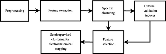
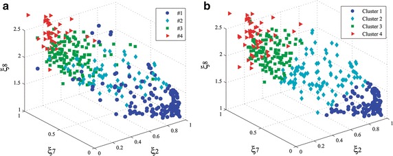
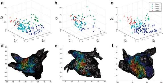
Methods: This work proposes a semi-supervised clustering method of four levels of fractionation. In particular, we make use of the spectral clustering that groups a set of widely used features extracted from atrial electrograms. We also introduce a new atrial-deflection-based feature to quantify the fractionated activity. Further, based on the sequential forward selection, we find the optimal subset that provides the highest performance in terms of the cluster validation. The method is tested on external validation of a labeled database. The generalization ability of the proposed training approach is tested to aid semi-supervised learning on unlabeled dataset associated with anatomical information recorded from three patients.
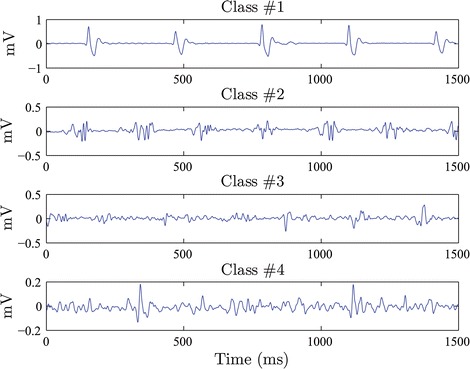
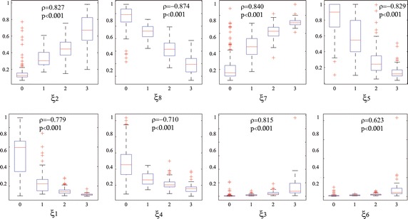
Results: A joint set of four extracted features, based on two time-domain morphology analysis and two non-linear dynamics, are selected. To discriminate between four considered levels of fractionation, validation on a labeled database performs a suitable accuracy (77.6 %). Results show a congruence value of internal validation index among tested patients that is enough to reconstruct the patterns over the atria to located critical sites with the benefit of avoiding previous manual classification of AF types.
Conclusions: To the best knowledge of the authors, this is the first work reporting semi-supervised clustering for distinguishing patterns in fractionated electrograms. The proposed methodology provides high performance for the detection of unknown patterns associated with critical EGM morphologies. Particularly, obtained results of semi-supervised training show the advantage of demanding fewer labeled data and less training time without significantly compromising accuracy. This paper introduces a new method, providing an objective scheme that enables electro-physiologist to recognize the diverse EGM morphologies reliably.
Keywords: Atrial fibrillation; Electrogram-guided ablation; Feature extraction; Spectral clustering.
Figures
References
Publication types
MeSH terms
LinkOut - more resources
Full Text Sources
Other Literature Sources
Molecular Biology Databases
