. 2016 Apr 27:6:25094.
doi: 10.1038/srep25094.

Terminalia Chebula provides protection against dual modes of necroptotic and apoptotic cell death upon death receptor ligation

Affiliations

Terminalia Chebula provides protection against dual modes of necroptotic and apoptotic cell death upon death receptor ligation

Yoonjung Lee et al. Sci Rep. .

Abstract

Death receptor (DR) ligation elicits two different modes of cell death (necroptosis and apoptosis) depending on the cellular context. By screening a plant extract library from cells undergoing necroptosis or apoptosis, we identified a water extract of Terminalia chebula (WETC) as a novel and potent dual inhibitor of DR-mediated cell death. Investigation of the underlying mechanisms of its anti-necroptotic and anti-apoptotic action revealed that WETC or its constituents (e.g., gallic acid) protected against tumor necrosis factor-induced necroptosis via the suppression of TNF-induced ROS without affecting the upstream signaling events. Surprisingly, WETC also provided protection against DR-mediated apoptosis by inhibition of the caspase cascade. Furthermore, it activated the autophagy pathway via suppression of mTOR. Of the WETC constituents, punicalagin and geraniin appeared to possess the most potent anti-apoptotic and autophagy activation effect. Importantly, blockage of autophagy with pharmacological inhibitors or genetic silencing of Atg5 selectively abolished the anti-apoptotic function of WETC. These results suggest that WETC protects against dual modes of cell death upon DR ligation. Therefore, WETC might serve as a potential treatment for diseases characterized by aberrantly sensitized apoptotic or non-apoptotic signaling cascades.


Figures

Figure 1
Figure 1. Chemical composition analysis of WETC determined using HPLC and UHPLC-HRMS analysis.
HPLC chromatograms of a mixture of six reference compounds (gallic acid, punicalagin, geraniin, chebulic acid, chebulagic acid and chebulinic acid) (A) and WETC (B) monitored at 276 nm. (C) Mass spectra of six reference compounds by UHPLC-HRMS. Gallic acid (m/z 169.0105 [M-H], TIC mode); Punicalagin (m/z 541.0281 [M-2H]2− z = 2, MRM mode); Geraniin (m/z 951.0754 [M-H], MRM mode); Chebulic acid (m/z 355.0308 [M-H], MRM mode); Chebulagic acid (m/z 953.0901 [M-H], MRM mode); Chebulinic acid (m/z 955.1058 [M-H], MRM mode).
Figure 2
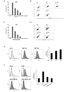
Figure 2. WETC protects TNF-induced necroptotic cell death by suppressing ROS production.
(A) L929 cells were pretreated with indicated concentrations of WETC for 30 min, followed by TNF (15 ng/mL) for another 8 h. Cells were trypsinized and collected in PBS, and cell death was quantified by trypan blue exclusion assay. Data were normalized to the rate of spontaneous cell death occurring in untreated cells. Data represent the mean ± SE of three independent experiments. *P < 0.05, compared with TNF-treated group. (B) L929 cells were pretreated with a pancaspase inhibitor (z-VAD-FMK, 20 μM), a necroptosis inhibitor (Nec-1, necrostatin-1, 10 μM) and WETC (0.4 mg/mL), followed by TNF (15 ng/mL) for another 8 h. The cells were stained with FITC-labeled annexin V and PI, and then analyzed by flow cytometry. (C) MEFs were pretreated with indicated concentrations of WETC for 30 min, and then treated with z-VAD-FMK (20 μM), TNF (15 ng/mL) and cycloheximide (CHX, 10 μg/mL) for 18 h. Cell death was quantified as in (A). *P < 0.05, compared with TNF/CHX/z-VAD-FMK-treated group. (D) MEFs were pretreated with WETC (0.4 mg/mL) and Nec-1 (10 μM), and then treated with TNF/CHX/z-VAD-FMK as in C. The mode of cell death was assessed as in (B). (E,F) L929 cells were treated with TNF (15 ng/mL) in the absence or presence of WETC (0.4 mg/mL) or mitochondria-targeted antioxidant Mito-TEMPO (100 μM) for various times, as indicated. The levels of intracellular ROS were monitored using the cell permeable dye, CM-H2DCFDA as described in Methods. Left panel, representative flow cytometry data. Right panel, quantification of ROS from three independent experiments.
Figure 3
Figure 3. WETC does not affect the upstream signaling complex formation of TNFR1.
L929 cells were treated with TNF (15 ng/mL) for 5 or 15 min in the absence or presence of WETC (0.4 mg/mL). Cell extracts from each sample were subjected to immunoprecipitation with anti-TNFR1 (A) and anti-IKK-γ (B) antibodies. Immunoprecipitates were analyzed by immunoblotting with indicated antibodies. One percent of cell extract from each treated sample was used as a control for protein content (Input).
Figure 4
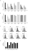
Figure 4. Inhibitory effects of the constituents of WETC on TNF-induced necroptotic cell death and ROS production.
(A,B) L929 cells were pretreated with WETC or indicated constituents for 30 min, followed by H2O2 ((A), 500 μM); or TNF ((B), 15 ng/mL) for another 8 h. Cell death was quantified as in Fig. 2A. Data represent the mean ± SE of three independent experiments. *P < 0.05, compared with H2O2-treated group. #P < 0.05, compared with TNF-treated group. (C,D) L929 cells were treated with TNF (15 ng/mL) in the absence or presence of indicated compounds for 6 h. The levels of intracellular ROS were monitored as in Fig. 2E. Representative flow cytometry data (C). Quantification of ROS from three independent experiments (D). GA: gallic acid, CA: chebulic acid, CGA: chebulagic acid, CNA: chebulinic acid, PU: punicalagin, GN: geraniin.
Figure 5
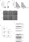
Figure 5. WETC protects DR-mediated apoptotic cell death in response to TNF and TRAIL.
(A) HeLa cells were pretreated with z-VAD-FMK (20 μM), and then treated with TNF (15 ng/mL) plus CHX (10 μg/mL) or TRAIL (500 ng/mL) for 8 h. Cell death was quantified as in Fig. 2A. (B) HeLa cells were pretreated with indicated concentrations of WETC for 30 min, followed by TNF/CHX or TRAIL, and cell death was quantified as in (A). *P < 0.05, compared with TNF/CHX-treated group. #P < 0.05, compared with TRAIL-treated group. (C,D) HeLa cells were treated with TNF/CHX or TRAIL in the absence or presence of WETC (0.4 mg/mL) for 8 h. Cell were visualized using a normal inverted microscope (C) and the mode of cell death (D) was assessed as in Fig. 2B (E,F). After pretreatment with WETC (0.4 mg/mL) for 30 min, HeLa cells were treated with TNF plus CHX or TRAIL for indicated times. Whole cell lysates were separated by SDS-PAGE and the immunoblotting was performed with indicated antibodies.
Figure 6
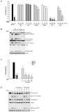
Figure 6. Inhibitory effects of the constituents of WETC on TRAIL-induced apoptotic cell death.
(A) HeLa cells were pretreated with WETC or indicated constituents for 30 min, followed by TRAIL (500 ng/mL) for another 8 h, and cell death was quantified as in Fig. 2A. *P < 0.05, compared with TRAIL-treated group. (B) After pretreatment with WETC (0.4 mg/mL) or each WETC constituent (40 μg/mL) for 30 min, HeLa cells were treated with TRAIL (500 ng/mL) for another 8 h. Whole cell lysates were separated by SDS-PAGE and the immunoblotting was performed with indicated antibodies. (C) HeLa cells were pretreated with WETC (0.4 mg/mL) or indicated concentrations of punicalagin (PU) for 30 min, followed by TRAIL (500 ng/mL) for another 8 h, and cell death was quantified as in (A). *P < 0.05, compared with TRAIL-treated group. (D) HeLa cells were pretreated with indicated concentrations of punicalagin (PU) or geraniin (GN) for 30 min, followed by TRAIL (500 ng/mL) for another 8 h. Whole cell lysates were immunoblotted with indicated antibodies as in (B).
Figure 7
Figure 7. WETC and its constituents (punicalagin and geraniin) induces autophagic flux via mTOR pathway.
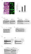
(A) HeLa cells were infected with recombinant adeno-viral expressing GFP-LC3 (100 PFU/cells) for 2 h, and then replaced with DMEM medium. After 24 h, cells were treated with WETC (0.4 mg/mL) and rapamycin (100 nM) for 6 h. Left panel, representative fluorescent images of the cells under a fluorescence microscope. Right panel, the percentage of cells with GFP-LC3 localized to punctated structures was calculated by counting a minimum of 100 cells per sample with values representing the means of triplicate experiments. (B) HeLa cells and MEFs were treated with the indicated concentrations (left panels) or times (right panels) of WETC. (C) WETC increases autophagic flux. After 30 min pretreatment with bafilomycin A (Baf-A, 100 nM), HeLa cells were further treated with WETC (0.4 mg/mL) for indicated times. (D) HeLa cells were treated with WETC (0.4 mg/mL) or each WETC constituent (40 μg/mL) for 6h. (E) HeLa cells and MEFs were treated with WETC for indicated times. (F) HeLa cells were treated with punicalagin (PU, 5 μg/mL) or geraniin (GN, 40 μg/mL) for indicated times. Whole cell lysates were separated by SDS-PAGE and the immunoblotting was performed with indicated antibodies.
Figure 8
Figure 8. Pharmacological or genetic inhibition of autophagy abolishes the anti-apoptotic potential of WETC.
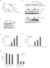
(A) HeLa cells were pretreated with autophagy inhibitor, NH4Cl (5 mM) and Baf-A (100 nM) for 30 min, and then followed by 500 ng/mL of TRAIL in the absence or presence of WETC (0.4 mg/mL) as indicated times. Cell death was quantified as in Fig. 2A. (B) HeLa cells were pretreated with NH4Cl and Baf-A, and then followed by TRAIL in the absence or presence of WETC for 6 h. Whole cell lysates were separated by SDS-PAGE and the immunoblotting was performed with indicated antibodies. (C) Tet-off Atg5 MEFs were incubated with or without 10 ng/mL doxycyclin hydrochloride (Dox) for 3 d, and then further treated with WETC (0.4 mg/mL) or TNF (15 ng/mL) plus CHX (10 μg/mL) for 6 h. Whole cell lysates were immunoblotted with indicated antibodies. (D,E) Tet-off Atg5 MEFs were treated with TNF plus CHX (T/C) or z-VAD-FMK, TNF and CHX (T/C/Z) in the absence or presence of Dox (10 ng/mL). (F) Tet-off Atg5 MEFs were pretreated with WETC (0.4 mg/mL) or punicalagin (PU, 5 μg/mL) for 30 min, and then followed by T/C or T/C/Z treatment for 18 h. The percentage of cells death was quantified as in A. Data represent the mean ± SE of three independent experiments. *P < 0.05, compared with TNF plus CHX-treated group.

Similar articles

Cited by

References

    1. Baud V. & Karin M. Signal transduction by tumor necrosis factor and its relatives. Trends Cell Biol 11, 372–377 (2001). - PubMed
    1. Wallach D. Cell death induction by TNF: a matter of self control. Trends Biochem Sci 22, 107–109 (1997). - PubMed
    1. Pennarun B. et al. Playing the DISC: turning on TRAIL death receptor-mediated apoptosis in cancer. Biochim Biophys Acta 1805, 123–140 (2010). - PubMed
    1. Guicciardi M. E. & Gores G. J. Life and death by death receptors. FASEB J 23, 1625–1637 (2009). - PMC - PubMed
    1. Gaur U. & Aggarwal B. B. Regulation of proliferation, survival and apoptosis by members of the TNF superfamily. Biochem Pharmacol 66, 1403–1408 (2003). - PubMed

Publication types

MeSH terms

LinkOut - more resources