Review
. 2016:74:103-41.
doi: 10.1016/bs.acc.2015.12.005. Epub 2016 Apr 7.

Tumor-Derived Exosomes and Their Role in Cancer Progression

Affiliations
Review

Tumor-Derived Exosomes and Their Role in Cancer Progression

Theresa L Whiteside. Adv Clin Chem. 2016.

Abstract

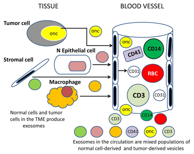
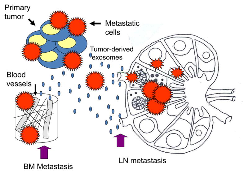
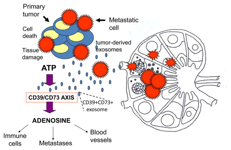
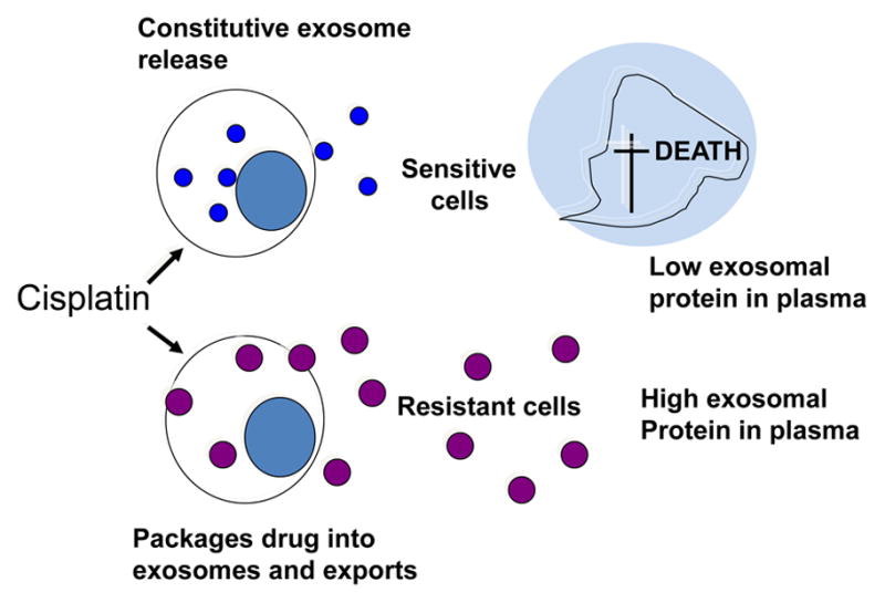
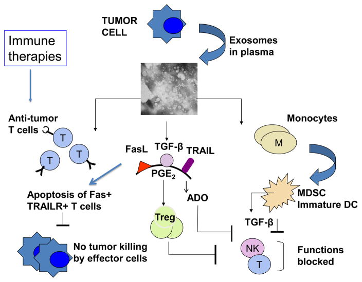
Tumor cells actively produce, release, and utilize exosomes to promote tumor growth. Mechanisms through which tumor-derived exosomes subserve the tumor are under intense investigation. These exosomes are information carriers, conveying molecular and genetic messages from tumor cells to normal or other abnormal cells residing at close or distant sites. Tumor-derived exosomes are found in all body fluids. Upon contact with target cells, they alter phenotypic and functional attributes of recipients, reprogramming them into active contributors to angiogenesis, thrombosis, metastasis, and immunosuppression. Exosomes produced by tumors carry cargos that in part mimic contents of parent cells and are of potential interest as noninvasive biomarkers of cancer. Their role in inhibiting the host antitumor responses and in mediating drug resistance is important for cancer therapy. Tumor-derived exosomes may interfere with cancer immunotherapy, but they also could serve as adjuvants and antigenic components of antitumor vaccines. Their biological roles in cancer development or progression as well as cancer therapy suggest that tumor-derived exosomes are critical components of oncogenic transformation.

Keywords: Cancer progression; Exosome content; Exosomes; Exosomes in immunoregulation; Information transfer; Tumor microenvironment; Tumor-derived exosomes.


Conflict of interest statement

Conflict of Interest

The author has no conflict of interest.

Figures

Figure 1
Figure 1
Exosome biogenesis and their release from a parent cell. The surface membrane undergoes endocytosis enclosing surface-residing molecules into an early endosome. Invagination of the endosomal membrane in late endosomes leads to the formation of intraluminal vesicles, each decorated with molecules originally present on the cell surface. Endosomes form a multivesicular body (MVB) containing pools of intraluminal vesicles. Fusion of the MVB with the cell surface membrane leads to release of free exosomes into the extracellular space. Note that components of the parent cell surface are now present on the membrane surrounding each exosome.
Figure 2
Figure 2
Exosome morphology and the molecular content. In A, transmission electron microscopy (TEM) of tumor-derived exosomes isolated from a tumor cell supernatant, pelleted by ultracentrifugation, embedded in Epon, sectioned and viewed by TEM. Note different sizes of membranous vesicles (courtesy of Dr. Simon Watkins, University of Pittsburgh). In B, the cartoon of an exosome illustrating the presence of a broad variety of molecules on its surface (some of the most common are indicated) and in the vesicle lumen.
Figure 3
Figure 3
Exosome uptake by cells. Exosomes may interact with target cells by one of several pathways, which depend on the cargo components they carry and on the presence of the recognizable “address” on the target cell membrane. Following the surface contact or uptake, exosomes transfer proteins and genes to the target cell and re-program its activities.
Figure 4
Figure 4
Tumor-derived exosomes and their local and systemic effects. Exosomes alter the tumor microenvironment (TME) and prepare distant tissue sites for metastasis. In the TME, exosomes interact with all cellular components and impact on the stromal, immune as well as vascular compartments. They access tumor-draining lymph nodes, facilitating entry of metastatic cells. They also carry and transfer oncogenes and oncogenic proteins to metastasizing tumor cells. Systemic effects of exosomes at distant tumor sites require that exosomes migrate via the blood or lymph. Once in place, they prepare tissue sites for metastasis or alter/educate the bone marrow (BM) environment, creating a pre-metastatic niche to promote tumor invasion and growth. They re-program mesenchymal stem cells (MSC) as well as progenitor hematopoietic cells. Tumor-derived exosomes interact with vascular endothelial cells (EC) favoring their proliferation, sprouting and the angiotube formation.
Figure 5
Figure 5
Exosomes present in the circulation or in body fluids of cancer patients are derived from many different normal and abnormal cells. Exosomes released into the extracellular space are distributed via the blood and lymph to distant body sites. In plasma, for example, tumor-derived exosomes represent only a small fraction of total exosome population which derives from normal circulating as well as tissue cells.
Figure 6
Figure 6
Pre-conditioning of lymph nodes (LN) or bone marrow (BM) niches by tumor-derived exosomes. These exosomes reach LNs or the BM prior to metastasizing tumor cells and re-program the local environment to facilitate the entry and “prepare the soil” for tumor cells.
Figure 7
Figure 7
Adenosine as a known metastasis-promoting factor. Tumor-derived exosomes carry CD39 and CD73, the ectonucleotidases which catalyze adenosine production in the presence of ATP. The TME is rich in ATP, and the activated CD39/CD73 axis promotes adenosine production. Adenosine induces inhibition of antitumor immune cell functions, promotes angiogenesis and favors metastasis, adenosine is only one of numerous other pro-tumorigenic factors that are regulated by tumor-derived exosomes.
Figure 8
Figure 8
Tumor-derived exosomes in drug resistance. Resistance of tumor cells to chemotherapy may be mediated by exosomes. Cisplatin-sensitive tumor cells die and, as a result, levels of tumor-derived exosomes decrease in plasma. In contrast, cisplatin-resistant tumor cells package the drug into exosomes and export large quantities of exosomes out of the cell. Thus, the exosomal content of plasma increases.
Figure 9
Figure 9
Systemic effects of tumor-derived exosomes on the host immune system. These effects may be direct, such as apoptosis of activated CD8+ T cells in the presence of FasL+ exosomes and blocking of monocyte differentiation into dendritic cells (DC), or indirect, such as exosome-driven expansion of regulatory T cells (Treg) or myeloid derived suppressor cells (MDSC) which suppress activities of other immune cells. Anti-tumor effector T cells generated in response to immune therapies are especially sensitive to exosomes carrying immunosuppressive factors. Natural killer (NK) cell activities are inhibited by transforming growth factor β (TGF-β) carried by exosomes or produced by MDSC. Prostaglandin2 (PGE2) and adenosine (ADO) are factors that are delivered by exosomes (PGE2) or produced by exosomes equipped with CD39/CD73 in the presence of ATP (ADO). The figure is reproduced with permission from ref (#39).

References

    1. Keller S, Ridinger J, Rupp AK, Janssen JW, Altevogt P. Body fluid derived exosomes as a novel template for clinical diagnostics. J Transl Med. 2011;9:86. - PMC - PubMed
    1. Brinton LT, Sloane HS, Kester M, Kelly KA. Formation and role of exosomes in cancer. Cell Mol Life Sci. 2015;72:659–71. - PMC - PubMed
    1. Gould SJ, Raposo G. As we wait: coping with an imperfect nomenclature for extracellular vesicles. J Extracell Vesicles. 2013;2 - PMC - PubMed
    1. van der Pol E, Boing AN, Harrison P, Sturk A, Nieuwland R. Classification, functions, and clinical relevance of extracellular vesicles. Pharmacol Rev. 2012;64:676–705. - PubMed
    1. Wilson DH, Rissin DM, Kan CW, Fournier DR, Piech T, Campbell TG, et al. The Simoa HD-1 Analyzer: A Novel Fully Automated Digital Immunoassay Analyzer with Single-Molecule Sensitivity and Multiplexing. J Lab Autom. 2015 - PubMed

Publication types

MeSH terms