. 2016 May 10;6(5):660-667.
doi: 10.1016/j.stemcr.2016.03.007. Epub 2016 Apr 21.

Basal Progenitors Contribute to Repair of the Prostate Epithelium Following Induced Luminal Anoikis

Affiliations

Basal Progenitors Contribute to Repair of the Prostate Epithelium Following Induced Luminal Anoikis

Roxanne Toivanen et al. Stem Cell Reports. .

Abstract

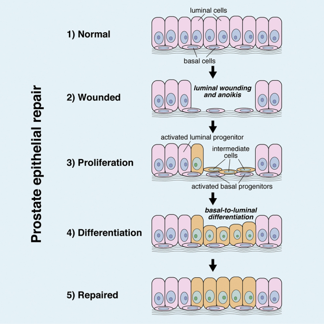
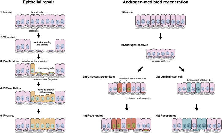
Contact with the extracellular matrix is essential for maintenance of epithelial cells in many tissues, while in its absence epithelial cells can detach and undergo anoikis. Here, we show that anoikis of luminal cells in the prostate epithelium is followed by a program of tissue repair that is mediated in part by differentiation of basal epithelial cells to luminal cells. We describe a mouse model in which inducible deletion of E-cadherin in prostate luminal cells results in their apoptotic cell death by anoikis, in the absence of phenotypic effects in the surrounding stroma. Quantitative assessments of proliferation and cell death in the luminal and basal compartments indicate that basal cells can rapidly generate luminal cells. Thus, our findings identify a role for basal-to-luminal differentiation in prostate epithelial repair, and provide a normal context to analogous processes that may occur during prostate cancer initiation.

Keywords: anoikis; epithelial lineage; prostate; stem cells; tissue repair.


Figures

None
Graphical abstract
Figure 1
Figure 1
Inducible Deletion of E-Cadherin in the Prostate Epithelium (A) Schematic timeline of the experiment. (B–G) H&E staining of histological sections from the anterior prostate. Arrows in (C) show patches of atypical cells, and arrows in (D), (E), and inset in (E) show cells sloughing into the lumen in Cdh1del prostates. (H–M) Immunofluorescence staining for E-cadherin and YFP. Arrows in (H) show intact E-cadherin expression in control mice; arrows in (I), (J), (K), and (M) show E-cadherin loss in YFP+ cells of Cdh1del prostates; and arrows in (L) indicate rare YFP+ cells in which E-cadherin was not deleted. (N–S) Immunofluorescence staining for p120 catenin and YFP. Arrows in (O), (P), and (Q) show cytoplasmic p120 staining in YFP+ cells of Cdh1del prostates. Numbers of mice examined: n = 7 for (B), (E), (H), (K), (N), and (Q); n = 5 for (C), (I), and (O); n = 4 for (D), (F), (G), (J), (L), (M), (P), (R), and (S). Scale bars, 50 μm. See also Figures S1 and S2; Tables S1 and S2.
Figure 2
Figure 2
Deletion of E-Cadherin in Prostate Luminal Cells Results in Anoikis without Reactive Stroma (A–C) Immunofluorescence staining of anterior prostate for CK18, E-cadherin, and YFP; arrows show CK18 expression in YFP+ cells. (D–F) Immunofluorescence staining for CK5, E-cadherin, and YFP; arrows show that YFP+ cells do not express CK5. (G–I) Immunofluorescence staining for CK18 and cleaved caspase-3 (CC3). The arrow in (H) shows co-localization of CC3 and CK18 in a sloughed cell; inset in (H) shows co-localization of CC3 (red) and YFP (green). (J–L) Immunohistochemical staining for smooth muscle α-actin shows normal appearance of smooth muscle in control and Cdh1del prostates (arrows). (M–O) Immunohistochemical staining for vimentin shows nearly undetectable expression in control and Cdh1del prostates. Numbers of mice examined: n = 3 for (A), (C), (D), (F), (G), (I), (J), (L), (M), and (O); n = 4 for (B), (E), (H), (K), and (N). Scale bars, 50 μm. See also Figure S3 and Table S3.
Figure 3
Figure 3
Basal Cells Contribute to Epithelial Repair following E-Cadherin Deletion (A–F) Immunofluorescence staining for CK5 and CK8 identifies double-positive intermediate cells (arrows). (G–L) Immunofluorescence staining for CK8, CK5, and Ki67 shows low levels of proliferation in luminal (arrows) and basal (arrowheads) cells of control and Cdh1del prostates. (M–O) Quantitation of the proportions of basal (M), luminal (N), and intermediate (O) cells in control and Cdh1del prostates. Values represent the mean and SD of at least three animals per group. (P and Q) Quantitation of cell proliferation (P) and cell death (Q) in the basal and luminal compartments. Values represent the mean and SD (error bars) of at least three animals per group. Numbers of mice examined: n = 4 for (A), (B), (C), (E), (G), (H), (I), and (K); n = 5 for (D) and (J); n = 3 for (F) and (L). Scale bars, 50 μm. See also Tables S4 and S5.
Figure 4
Figure 4
Assay Dependence of Stem/Progenitor Activity in the Prostate Epithelium Schematic depiction of tissue repair and androgen-mediation regeneration of the prostate epithelium; newly generated cells are indicated by different colors. During tissue repair, luminal epithelial cells are primarily generated by a process of basal proliferation and basal-to-luminal differentiation in which “intermediate” cells represent a transitional state; in addition, there is activity of unipotent luminal progenitors. During androgen-mediated regeneration, tissue growth is largely mediated by unipotent luminal and basal progenitors, with a significant contribution by bipotential luminal stem cells (castration-resistant Nkx3.1-expressing cells [CARNs]).

References

    1. Blanpain C., Fuchs E. Stem cell plasticity. Plasticity of epithelial stem cells in tissue regeneration. Science. 2014;344:1242281. - PMC - PubMed
    1. Bonkhoff H., Remberger K. Differentiation pathways and histogenetic aspects of normal and abnormal prostatic growth: a stem cell model. Prostate. 1996;28:98–106. - PubMed
    1. Boussadia O., Kutsch S., Hierholzer A., Delmas V., Kemler R. E-cadherin is a survival factor for the lactating mouse mammary gland. Mech. Dev. 2002;115:53–62. - PubMed
    1. Burger P.E., Xiong X., Coetzee S., Salm S.N., Moscatelli D., Goto K., Wilson E.L. Sca-1 expression identifies stem cells in the proximal region of prostatic ducts with high capacity to reconstitute prostatic tissue. Proc. Natl. Acad. Sci. USA. 2005;102:7180–7185. - PMC - PubMed
    1. Chiarugi P., Giannoni E. Anoikis: a necessary death program for anchorage-dependent cells. Biochem. Pharmacol. 2008;76:1352–1364. - PubMed

Publication types

LinkOut - more resources