. 2016 Apr 26:9:237.
doi: 10.1186/s13071-016-1511-8.

Transmission mechanisms of an emerging insect-borne rickettsial pathogen

Affiliations

Transmission mechanisms of an emerging insect-borne rickettsial pathogen

Lisa D Brown et al. Parasit Vectors. .

Abstract

Background: Vector-borne pathogens must overcome arthropod infection and escape barriers (e.g. midgut and salivary glands) during the extrinsic incubation period (EIP) before subsequent transmission to another host. This particular timespan is undetermined for the etiological agent of flea-borne spotted fever (Rickettsia felis). Artificial acquisition of R. felis by blood-feeding cat fleas revealed dissemination to the salivary glands after seven days; however, this length of time is inconsistent with co-feeding studies that produced infectious cat fleas within 24 h of infection. In the current study, we demonstrated that an alternative mechanism is responsible for the early-phase transmission that typifies flea-borne R. felis spread.

Methods: Co-feeding transmission bioassays were constructed to assess temporal dynamics of R. felis amongst cat fleas, including exposure time to produce infectious fleas and association time to transmit infection to naïve fleas. Additional experiments examined the proportion of R. felis-exposed cat fleas with contaminated mouthparts, as well as the likelihood for cat fleas to release R. felis from their mouthparts following exposure to an infectious bloodmeal. The potential for mechanical transmission of R. felis by co-feeding cat fleas was further examined using fluorescent latex beads, as opposed to a live pathogen, which would not require a biological mechanism to achieve transmission.

Results: Analyses revealed that R. felis-infected cat fleas were infectious to naïve fleas less than 24 h after exposure to the pathogen, but showed no rickettsial dissemination to the salivary glands during this early-phase transmission. Additionally, the current study revealed that R. felis-infected cat fleas must co-feed with naïve fleas for more than 12 h in order for early-phase transmission to occur. Further evidence supported that contaminated flea mouthparts may be the source of the bacteria transmitted early, and demonstrated that R. felis is released from the mouthparts during brief probing events. Moreover, the use of fluorescent latex beads supports the notion that early-phase transmission of R. felis is a mechanical mechanism.

Conclusions: Determination of the transmission mechanisms utilized by R. felis is essential to fully understand the vulnerability of susceptible vertebrate hosts, including humans, to this pathogen.

Keywords: Cat fleas; Rickettsia felis; Transmission mechanisms.


Figures
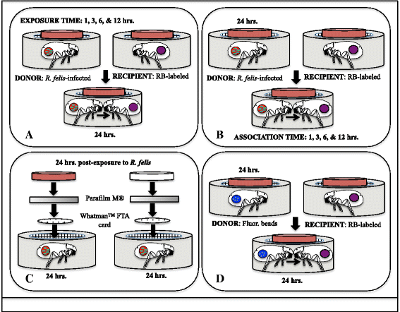
Fig. 1
Fig. 1
Diagrams of experimental designs. a Cat fleas were exposed to an infectious bloodmeal for 1, 3, 6 or 12 h, and then divided into feeding capsules containing naïve cat fleas for 24 h (exposure bioassays); b Cat fleas were exposed to an infectious bloodmeal for 24 h and then divided into feeding capsules containing naïve cat fleas for 1, 3, 6 or 12 h (association bioassays); c Whatman™ FTA cards were placed in flea cages after 24 hpe to an R. felis-infected bloodmeal. Cat fleas either had access to blood or the bloodmeal was removed for the duration of the experiment; d Cat fleas were exposed to an “infectious” bloodmeal containing fluorescent latex beads for 24 h, and then were placed with naïve fleas for 24 h
Fig. 2
Fig. 2
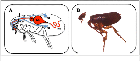
Flea dissections. a Diagram of flea internal anatomy. The dash line represents where the incision for dissections was made (PV,  proventriculus; MG, midgut; HG, hindgut; SG, salivary glands); b Photographic image of flea dissections to determine the presence of R. felis in flea mouthparts versus midgut at 24 hpe to an infectious bloodmeal
Fig. 3
Fig. 3
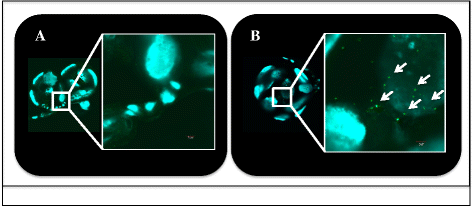
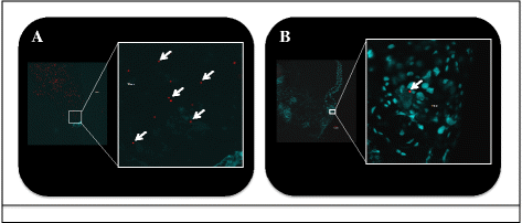
Dissemination of Rickettsia to flea salivary glands. a No rickettsial antigen is present at 1 dpe to an R. felis-infected bloodmeal; b Presence of rickettsial antigen (labeled green, indicated by arrows) at 28 dpe to an R. felis-infected bloodmeal (positive control)
Fig. 4
Fig. 4
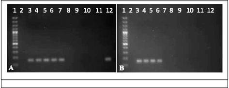
PCR detection of rickettsial 17-kDa antigen gene in Whatman™ FTA cards. a Lane 1, 100 bp DNA marker; Lane 2, blank; Lanes 3–7, single disc punch from five different cards exposed to R. felis-infected cat fleas in the presence of blood; Lanes 8–10, blank; Lane 11, environmental control; Lane 12, positive PCR R. felis genomic DNA; b Lane 1, 100 bp DNA marker; Lanes 2, 7–11, blank; Lanes 3, 4, 5, single disc punch from three different cards exposed to R. felis-infected, surface sterilized cats fleas in the absence of blood; Lane 6, positive PCR R. felis genomic DNA; Lane 12, environmental control
Fig. 5
Fig. 5
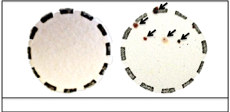
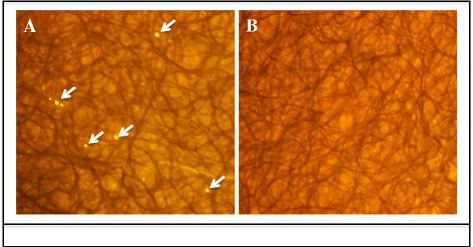
FTA cards exposed to cat fleas in the absence (left) and presence (right) of blood. Residual blood droplets (arrows) were deposited when cat fleas had access to blood
Fig. 6
Fig. 6
Dissections of cat flea midguts exposed to fluorescent latex beads. a Donor cat flea with fluorescent beads (arrows) after 1 day post-exposure to an “infectious” bloodmeal; b Recipient cat flea with fluorescent beads (arrow) after 1 day of co-feeding with donor cat fleas
Fig. 7
Fig. 7
Whatman™ FTA cards placed in cat flea cages at 24 hpe to fluorescent latex beads in blood. a Cat fleas deposited beads (arrows) onto cards following surface sterilization and no access to blood; b Whatman™ FTA card exposed to non-experimental cat fleas with no access to blood

References

    1. Richards AL, Jiang J, Omulo S, Dare R, Abdirahman K, Ali A, et al. Human infection with Rickettsia felis, Kenya. Emerg Infect Dis. 2010;16(7):1081–6. doi: 10.3201/eid1607.091885. - DOI - PMC - PubMed
    1. Socolovschi C, Mediannikov O, Sokhna C, Tall A, Diatta G, Bassene H, et al. Rickettsia felis-associated uneruptive fever, Senegal. Emerg Infect Dis. 2010;16(7):1140–2. doi: 10.3201/eid1607.100070. - DOI - PMC - PubMed
    1. Mediannikov O, Socolovschi C, Edouard S, Fenollar F, Mouffok N, Bassene H, et al. Common epidemiology of Rickettsia felis infection and malaria, Africa. Emerg Infect Dis. 2013;19(11):1775–83. doi: 10.3201/eid1911.130361. - DOI - PMC - PubMed
    1. Mediannikov O, Fenollar F, Bassene H, Tall A, Sokhna C, Trape JF, et al. Description of “yaaf”, the vesicular fever caused by acute Rickettsia felis infection in Senegal. J Infect. 2013;66(6):536–40. doi: 10.1016/j.jinf.2012.10.005. - DOI - PubMed
    1. Mediannikov O, Socolovschi C, Million M, Sokhna C, Bassene H, Diatta G, et al. Molecular identification of pathogenic bacteria in eschars from acute febrile patients, Senegal. Am J Trop Med Hyg. 2014;91(5):1015–9. doi: 10.4269/ajtmh.13-0629. - DOI - PMC - PubMed

Publication types

MeSH terms

LinkOut - more resources