Review
. 2016 Sep;7(5):620-36.
doi: 10.1002/wrna.1354. Epub 2016 Apr 27.

Structural insights into ribosome translocation

Affiliations
Review

Structural insights into ribosome translocation

Clarence Ling et al. Wiley Interdiscip Rev RNA. 2016 Sep.

Abstract

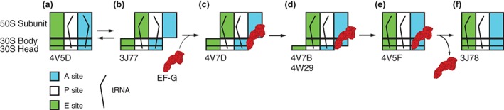
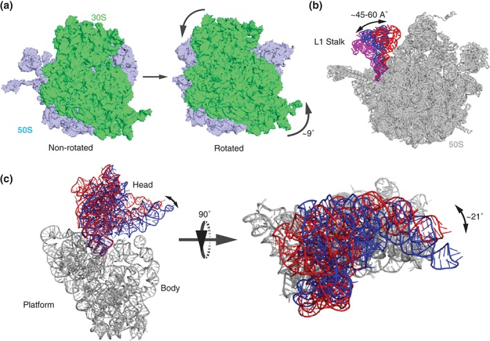
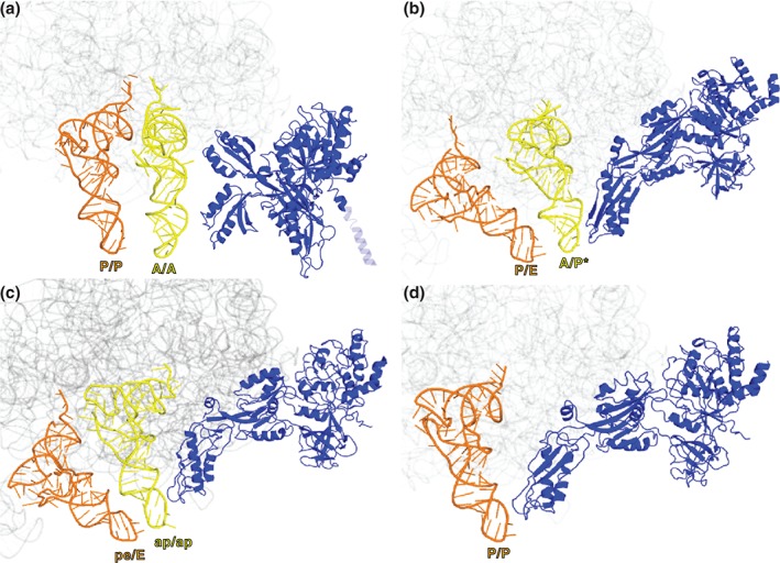
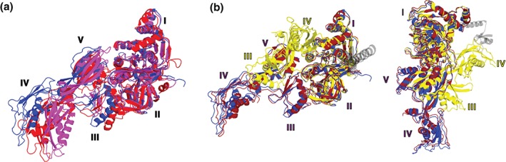
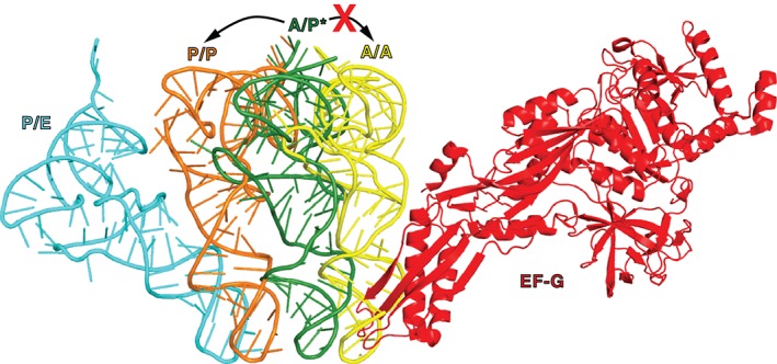
During protein synthesis, tRNA and mRNA are translocated from the A to P to E sites of the ribosome thus enabling the ribosome to translate one codon of mRNA after the other. Ribosome translocation along mRNA is induced by the universally conserved ribosome GTPase, elongation factor G (EF-G) in bacteria and elongation factor 2 (EF-2) in eukaryotes. Recent structural and single-molecule studies revealed that tRNA and mRNA translocation within the ribosome is accompanied by cyclic forward and reverse rotations between the large and small ribosomal subunits parallel to the plane of the intersubunit interface. In addition, during ribosome translocation, the 'head' domain of small ribosomal subunit undergoes forward- and back-swiveling motions relative to the rest of the small ribosomal subunit around the axis that is orthogonal to the axis of intersubunit rotation. tRNA/mRNA translocation is also coupled to the docking of domain IV of EF-G into the A site of the small ribosomal subunit that converts the thermally driven motions of the ribosome and tRNA into the forward translocation of tRNA/mRNA inside the ribosome. Despite recent and enormous progress made in the understanding of the molecular mechanism of ribosome translocation, the sequence of structural rearrangements of the ribosome, EF-G and tRNA during translocation is still not fully established and awaits further investigation. WIREs RNA 2016, 7:620-636. doi: 10.1002/wrna.1354 For further resources related to this article, please visit the WIREs website.


Figures

Figure 1
Figure 1
Structural organization of the ribosome and elongation factor G. (a) Crystal structure of the 70S ribosome (Protein Data Bank ID [PDBID] 4V6F1). Large, 50S subunit and small, 30S subunit are colored in light blue and light green, respectively. A‐site, P‐site, and E‐site tRNAs are shown in yellow, orange, and red, respectively. mRNA is colored purple. A box diagram of the ribosome showing tRNAs bound in the A, P, and E sites of the 50S and 30S subunits is shown below the crystal structure of the 70S ribosome. (b) Crystal structure of ribosome‐free EF‐G (PDBID 1DAR2) with domains color‐coded: G′ domain (dark blue), G domain (green), domain II (dark red), domain III (orange), domain IV (magenta), and domain V (light blue).
Figure 2
Figure 2
Scheme of tRNA rearrangements during EF‐G‐catalyzed ribosome translocation. Diagrams show tRNA positions relative to the A, P, and E sites on the 50S subunit and 30S head and body. (a) peptidyl‐ and deacylated tRNAs are bound in A/A and P/P classical states, (b) A/P and P/E hybrid states, (c) A/P* and P/E states in the presence of ribosome‐bound EF‐G, (d) ap/P and pe/E chimeric states in the presence of ribosome‐bound EF‐G, (e) classical P/P and E/E state in the presence of ribosome‐bound EF‐G, and (f) classical P/P and E/E state after EF‐G dissociation. Please see additional details in the text. PDB IDs corresponding to each structural state are indicated under the schematic.
Figure 3
Figure 3
Structural rearrangements of the ribosome that accompany translocation. (a) Structures of the ribosome in the non‐rotated, classical state54 (PDBID 4V9D) and the rotated, hybrid state55 (PDBID 4V7C). The small subunit is shown in light green and the large subunit is shown light blue. The ribosome is viewed from the solvent side of the small subunit. Curved arrows indicate the counter‐clockwise rotation of the small subunit relative to the large subunit. (b) 50S L1 stalk (23S rRNA helices 76, 77, and 78) is shown in the closed54 (red, PDBID 4V9D), half‐closed1 (blue, PDBID 4V6F), and open54 (magenta, PDBID 4V9D) positions. The rest of the large subunit is shown in gray. Structures were superimposed by structural alignment of 23S rRNA. (c) The swiveling motion of the head domain of the small subunit is shown by structural alignment of the body and platform domains of 16S rRNA (shown in gray) of crystal structures of nonrotated ribosome containing classical A/A, P/P, and E/E site tRNAs (head domain in blue, PDBID 4V5156) and partially rotated ribosome containing a chimeric ap/ap and pe/E tRNAs (head domain in red, PDBID 4W2942). The 30S is viewed from its solvent side (left) and the ‘top’ of the small subunit head (right). Double‐headed arrows indicate the direction of head swiveling, which is perpendicular to the long axis of the small subunit.
Figure 4
Figure 4
Structural rearrangements of EF‐G on the ribosome during translocation. (a) EF‐G (blue in all structures) bound to the nonrotated pretranslocation ribosome containing A/A‐ and P/P‐ tRNAs93 (PDBID 4WPO). The N‐terminal domain of large subunit protein L9 covalently linked to the N‐terminus of EF‐G is shown as a transparent blue. (b) EF‐G bound to the fully rotated pretranslocation ribosome containing A/P*‐ and P/E‐ tRNAs55 (PDBID 4V7D); (c) EF‐G bound to partially rotated ribosomes containing chimeric ap/ap and pe/E‐ tRNAs42 (PDBID 4 W29) and (d) EF‐G bound to the nonrotated posttranslocation ribosome containing P/P‐ and E/E‐ tRNAs13 (PDBID 4V5F). 23S rRNA in all structures is shown in gray.
Figure 5
Figure 5
Movement of EF‐G from the pre‐ to posttranslocation conformation on the ribosome. EF‐G (red, PDBID 4V7D55) bound to the viomycin‐trapped pretranslocation ribosome is superimposed with EF‐G bound to the posttranslocation ribosome (blue, PDBID 4V5F13). The superimposition is obtained by structural alignment of respective 23S rRNAs. A/P* tRNA is shown in yellow. 23S rRNA of pre and posttranslocation ribosomes are shown in transparent red and blue, respectively.
Figure 6
Figure 6
Interdomain rearrangements of EF‐G. (a) EF‐G in the posttranslocation conformation (blue, PDBID 4V5F13) is superimposed by structural alignment of domains I and II with ribosome‐free EF‐G•GDP (magenta, PDBID 1DAR2) and EF‐G bound to the viomycin‐trapped pretranslocation ribosome (red, PDBID 4V7D55), (b) EF‐G in the posttranslocation conformation (blue, PDBID 4V5F13) is superimposed by structural alignment of domains I and II with EF‐G bound to a ribosome containing a chimeric ap/ap tRNA (dark red, PDBID 4W2942) and EF‐G‐L9 fusion bound to the nonrotated, pretranslocation ribosome (yellow, PDBID 4WPO93). The N‐terminal domain of L9 covalently linked to EF‐G (PDBID 4WPO93) is shown in transparent gray. Two differently oriented views of EF‐G structures are shown. Domains of EF‐G are numbered as indicated.
Figure 7
Figure 7
EF‐G may promote tRNA translocation by clashing with A‐site tRNA. A/A (yellow, PDBID 4V5D107), A/P* (dark green, PDBID 4V7D55), P/P (orange, PDBID 4V5D107), and P/E (cyan, PDBID 4V7C55) tRNAs are superimposed by structural alignment of 23S rRNA in the respective structures. EF‐G bound to the viomycin‐trapped pretranslocation ribosome is shown in red (PDBID 4V7D55). In the absence of EF‐G, spontaneous intersubunit rotation is coupled to the fluctuation of tRNAs between classical (A/A and P/P) and hybrid (A/P [A/P*] and P/E) states. In the presence of ribosome‐bound EF‐G, upon the reverse intersubunit rotation from the rotated to nonrotated conformations of the ribosome, the movement of peptidyl‐tRNA from A/P* to A/A state is disallowed because of the clash with domain IV of EF‐G and, thus, the peptidyl‐tRNA translocates into the P/P state.

References

FURTHER READING

    1. Frank J, Gonzalez RL Jr. Structure and dynamics of a processive Brownian motor: the translating ribosome. Annu Rev Biochem 2010, 79:381–412. - PMC - PubMed
    1. Frank J. Molecular Machines in Biology: Workshop of the Cell. Cambridge and New York: Cambridge University Press; 2011.
    1. Moore PB. How should we think about the ribosome? Annu Rev Biophys 2012, 41:1–19. - PubMed
    1. Voorhees RM, Ramakrishnan V. Structural basis of the translational elongation cycle. Annu Rev Biochem 2013, 82:203–236. - PubMed

References

    1. Jenner LB, Demeshkina N, Yusupova G, Yusupov M. Structural aspects of messenger RNA reading frame maintenance by the ribosome. Nat Struct Mol Biol 2010, 17:555–560. - PubMed
    1. AEvarsson A, Brazhnikov E, Garber M, Zheltonosova J, Chirgadze Y, al‐Karadaghi S, Svensson LA, Liljas A. Three‐dimensional structure of the ribosomal translocase: elongation factor G from thermus thermophilus. EMBO J 1994, 13:3669–3677. - PMC - PubMed
    1. Czworkowski J, Wang J, Steitz TA, Moore PB. The crystal structure of elongation factor G complexed with GDP, at 2.7 A resolution. EMBO J 1994, 13:3661–3668. - PMC - PubMed
    1. Rodnina MV, Savelsbergh A, Katunin VI, Wintermeyer W. Hydrolysis of GTP by elongation factor G drives tRNA movement on the ribosome. Nature 1997, 385:37–41. - PubMed
    1. Katunin VI, Savelsbergh A, Rodnina MV, Wintermeyer W. Coupling of GTP hydrolysis by elongation factor G to translocation and factor recycling on the ribosome. Biochemistry 2002, 41:12806–12812. - PubMed

Publication types

MeSH terms

LinkOut - more resources