. 2016 Sep;44(9):866-873.e4.
doi: 10.1016/j.exphem.2016.04.010. Epub 2016 Apr 23.

Long-term adaptation to hypoxia preserves hematopoietic stem cell function

Affiliations

Long-term adaptation to hypoxia preserves hematopoietic stem cell function

Jichun Chen et al. Exp Hematol. 2016 Sep.

Abstract

Molecular oxygen sustains aerobic life, but it also serves as the substrate for oxidative stress, which has been associated with the pathogenesis of disease and with aging. Compared with mice housed in normoxia (21% O2), reducing ambient oxygen to 10% O2 (hypoxia) resulted in increased hematopoietic stem cell (HSC) function as measured by bone marrow (BM) cell engraftment onto lethally irradiated recipients. The number of BM c-Kit(+)Sca-1(+)Lin(-) (KSL) cells as well as the number of cells with other hematopoietic stem and progenitor cell markers were increased in hypoxia mice, whereas the BM cells' colony-forming capacity remained unchanged. KSL cells from hypoxia mice showed a decreased level of oxidative stress and increased expression of transcription factor Gata1 and cytokine receptor c-Mpl, consistent with the observations of increased erythropoiesis and enhanced HSC engraftment. These observations demonstrate the benefit of a hypoxic HSC niche and suggest that hypoxic conditions can be further optimized to preserve stem cell integrity in vivo.


Figures

Figure 1.
Figure 1.
Hypoxia increases BM HSC engraftment in vivo in B6-ApoE−/− mice. (A) BM cells from hypoxia (N=5) or normoxia (N=5) B6-ApoE−/− donors were mixed 1:1 with BM cells from a pool of B6-CD45.1 congenic animals. These cell mixtures were engrafted into lethally-irradiated (11 Gy TBI) B6-CD45.1 or normal B6 recipients at 2 × 10⁶ cells/recipient (3-5 recipients/donor) in a competitive repopulation assay. (B) Contribution of hypoxia donors to mature blood cells was detected at 1 to 9 mo after BM transplantation, P<0.05. (C) Hypoxia versus normoxia donor-derived KSL cells in BM 9 mo following transplantation. (D) Representative FACS profile of hypoxia versus normoxia donor BM cell reconstitution of peripheral blood CD3+ T cells, CD11b+ myeloid cells and CD45R+ B cells at 9 mo. Values shown as mean ± SE, *P<0.05
Figure 2.
Figure 2.
Hypoxia augments HSC function in wild-type B6 mice. (A) BM cells from hypoxia (N=5) and normoxia (N=5) donors were each mixed 1:1 with a pool of BM cells from B6-CD45.1 competitors and the BM mixture were injected into lethally-irradiated (11 Gy TBI) B6-CD45.1 recipients at 2 × 10⁵ donor and 2 × 10⁵ competitor BM cells per recipient, 2-3 recipient/donor. (B) Contribution of hypoxia donors to mature blood cells was measured at 1 to 5 mo after BM transplantation, P<0.01. (C) Donor contributions were calculated as repopulation unit (RUs/105 donor BM cells), P<0.01.
Figure 3.
Figure 3.
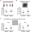
Hypoxia has no significant effect on BM colony formation. (A) BM cells from hypoxia (N=5) and normoxia (N=5) B6-ApoE−/− mice were tested for colony forming units-spleen (CFU-s) in vivo. BM cells from hypoxia and normoxia mice were transplanted into lethally irradiated (11 Gys) B6 recipients at 105 cells/recipient (3 recipients/donor) and splenic colonies counted at d 12. Representative splenic colonies are shown. (B) In vitro myeloid colony form cells (CFC) were measured using hypoxia and normoxia BM cells plated on methylcellulose media at 37°C with 5% CO2 for 12 d. (C) In vitro cobblestone area forming cell (CAFC) assay was performed using BM cells from hypoxia and normoxia donors overlaid on irradiated stromal feeder cells. CAFC was counted at 1 and 3 wk.
Figure 4.
Figure 4.
Hypoxia increases KSL and BM cellularity in association with decreased oxidative stress in B6-ApoE−/− mice. (A) Total BM cell counts under normoxia (N=10) or chronic hypoxia (N=10). (B) KSL cell counts in BM of normoxia versus hypoxia mice. (C) KSL cell frequency in BM of normoxia versus hypoxia mice by FACS profile. (D) BM cells were stained with DCF-DA as indicator of whole cell ROS level, shown as representative FACS profile and percentage ROS+ cells in BM KSL cells. (E) Gata1 and c-Mpl mRNA expression was measured in KSL cells from hypoxia (N=5-12) and normoxia (N=5-17) mice using real time RT-PCR. Values shown as mean ± SE, *P<0.05, **P<0.01.

Similar articles

Cited by

References

    1. West JB. Highest permanent human habitation. High Alt Med Biol. 2002; 3:401–407. - PubMed
    1. Mortimer EA Jr., Monson RR, MacMahon B. Reduction in mortality from coronary heart disease in men residing at high altitude. N Engl J Med. 1977; 296:581–585. - PubMed
    1. Weinberg CR, Brown KG, Hoel DG. Altitude, radiation, and mortality from cancer and heart disease. Radiat Res. 1987; 112:381–390. - PubMed
    1. Faeh D, Gutzwiller F, Bopp M. Lower mortality from coronary heart disease and stroke at higher altitudes in Switzerland. Circulation. 2009; 120:495–501. - PubMed
    1. Winkelmayer WC, Liu J, Brookhart MA. Altitude and all-cause mortality in incident dialysis patients. JAMA. 2009; 301:508–512. - PubMed

Publication types