

The tract terminations in the temporal lobe: Their location and associated functions
- PMID: 27118049
- PMCID: PMC5726606
- DOI: 10.1016/j.cortex.2016.03.013
The tract terminations in the temporal lobe: Their location and associated functions
Abstract
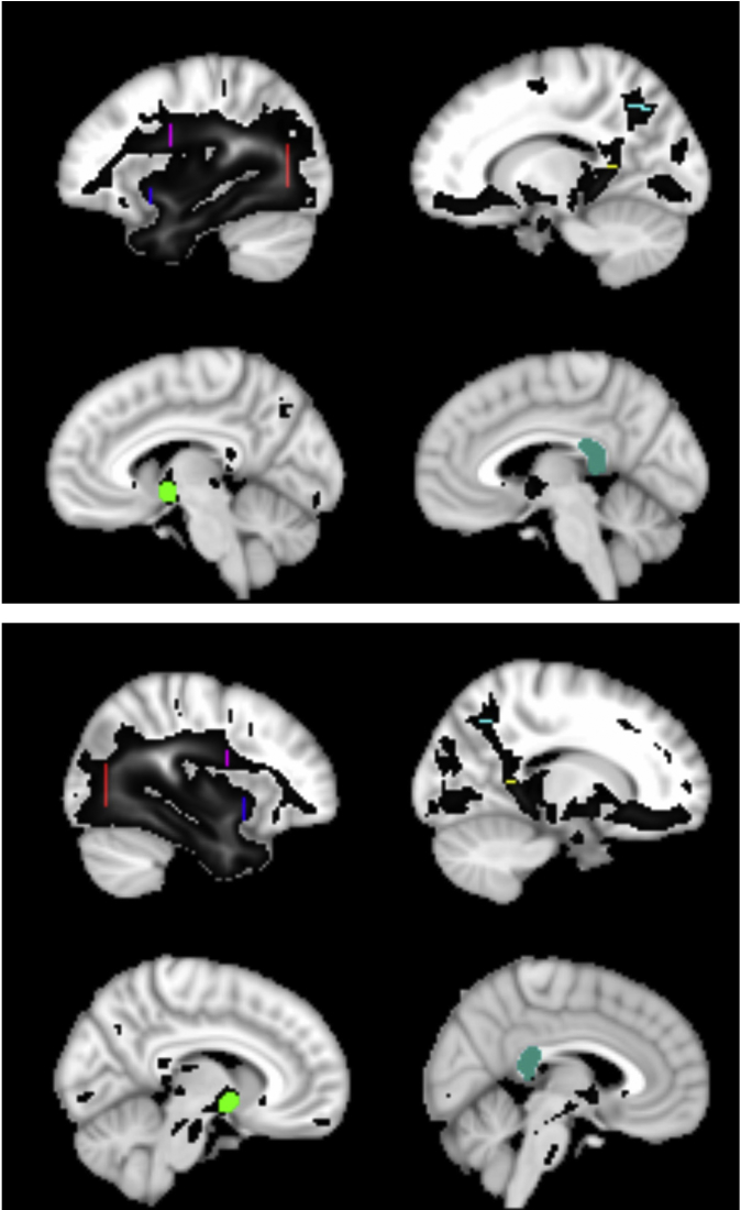
Temporal lobe networks are associated with multiple cognitive domains. Despite an upsurge of interest in connectional neuroanatomy, the terminations of the main fibre tracts in the human brain are yet to be mapped. This information is essential given that neurological, neuroanatomical and computational accounts expect neural functions to be strongly shaped by the pattern of white-matter connections. This paper uses a probabilistic tractography approach to identify the main cortical areas that contribute to the major temporal lobe tracts. In order to associate the tract terminations to known functional domains of the temporal lobe, eight automated meta-analyses were performed using the Neurosynth database. Overlaps between the functional regions highlighted by the meta-analyses and the termination maps were identified in order to investigate the functional importance of the tracts of the temporal lobe. The termination maps are made available in the Supplementary Materials of this article for use by researchers in the field.
Keywords: Brain mapping; Diffusion MRI; Temporal lobe; Tractography; White matter.
Copyright © 2016 The Authors. Published by Elsevier Ltd.. All rights reserved.
Figures
Similar articles
-
Revisiting the human uncinate fasciculus, its subcomponents and asymmetries with stem-based tractography and microdissection validation.Brain Struct Funct. 2017 May;222(4):1645-1662. doi: 10.1007/s00429-016-1298-6. Epub 2016 Aug 31. Brain Struct Funct. 2017. PMID: 27581617
-
Plasticity of left perisylvian white-matter tracts is associated with individual differences in math learning.Brain Struct Funct. 2016 Apr;221(3):1337-51. doi: 10.1007/s00429-014-0975-6. Epub 2015 Jan 21. Brain Struct Funct. 2016. PMID: 25604464 Free PMC article.
-
Mapping temporo-parietal and temporo-occipital cortico-cortical connections of the human middle longitudinal fascicle in subject-specific, probabilistic, and stereotaxic Talairach spaces.Brain Imaging Behav. 2017 Oct;11(5):1258-1277. doi: 10.1007/s11682-016-9589-3. Brain Imaging Behav. 2017. PMID: 27714552 Free PMC article.
-
The connectional anatomy of the temporal lobe.Handb Clin Neurol. 2022;187:3-16. doi: 10.1016/B978-0-12-823493-8.00001-8. Handb Clin Neurol. 2022. PMID: 35964979 Review.
-
Anatomy of the medial temporal lobe.Magn Reson Imaging. 1995;13(8):1047-55. doi: 10.1016/0730-725x(95)02012-i. Magn Reson Imaging. 1995. PMID: 8750316 Review.
Cited by
-
Unveiling the dynamic interplay between the hub- and spoke-components of the brain's semantic system and its impact on human behaviour.Neuroimage. 2019 Oct 1;199:114-126. doi: 10.1016/j.neuroimage.2019.05.059. Epub 2019 May 24. Neuroimage. 2019. PMID: 31132452 Free PMC article.
-
Directional changes in information flow between human brain cortical regions after application of anodal transcranial direct current stimulation (tDCS) over Broca's area.Biomed Opt Express. 2018 Oct 10;9(11):5296-5317. doi: 10.1364/BOE.9.005296. eCollection 2018 Nov 1. Biomed Opt Express. 2018. PMID: 30460129 Free PMC article.
-
Changes in brain structure in subjects with resistance to thyroid hormone due to THRB mutations.Thyroid Res. 2023 Nov 6;16(1):34. doi: 10.1186/s13044-023-00176-2. Thyroid Res. 2023. PMID: 37592301 Free PMC article.
-
The anesthetic approach for endovascular recanalization therapy depends on the lesion site in acute ischemic stroke.Neuroradiology. 2021 Dec;63(12):2121-2129. doi: 10.1007/s00234-021-02762-3. Epub 2021 Jul 10. Neuroradiology. 2021. PMID: 34244817 Free PMC article.
-
Distinct contributions of the fornix and inferior longitudinal fasciculus to episodic and semantic autobiographical memory.Cortex. 2017 Sep;94:1-14. doi: 10.1016/j.cortex.2017.05.010. Epub 2017 Jun 20. Cortex. 2017. PMID: 28710907 Free PMC article.
References
-
- Andersson J.L., Jenkinson M., Smith S. 2010. Non-linear registration, aka spatial normalisation. FMRIB technical report TR07JA2.
-
- Azadbakht H., Parkes L.M., Haroon H.A., Augath M., Logothetis N.K., De Crespigny A.J. Proceedings of the 20th Scientific Meeting and Exhibition of the International Society for Magnetic Resonance in Medicine. The International Society for Magnetic Resonance in Medicine; Melbourne, Australia: 2012. Validation of tractography against in vivo tracing in the macaque visual system – effect of distance correction.
-
- Bajada C.J., Lambon Ralph M.A., Cloutman L.L. Transport for Language South of the Sylvian Fissure: the routes and history of the main tracts and stations in the ventral language network. Cortex: A Journal Devoted To the Study of the Nervous System and Behavior. 2015;19(69):141–151. - PubMed
-
- Basser P.J. ISMRM; 1998. Fiber-tractography via diffusion tensor MRI (DT-MRI). Book of Abstracts: Sixth Annual Meeting of the International Society for Magnetic Resonance in Medicine; p. 1226.
Publication types
MeSH terms
Grants and funding
LinkOut - more resources
Full Text Sources
Other Literature Sources
