Review
. 2016 Aug;30(8):2640-50.
doi: 10.1096/fj.201500073R. Epub 2016 Apr 26.

CIB1: a small protein with big ambitions

Affiliations
Review

CIB1: a small protein with big ambitions

Tina M Leisner et al. FASEB J. 2016 Aug.

Abstract

Calcium- and integrin-binding protein 1 (CIB1) is a small, ubiquitously expressed protein that was first identified as an intracellular binding partner of a platelet-specific α-integrin cytoplasmic tail. Although early studies revealed a role for CIB1 in regulating platelet integrin activity, recent studies have indicated a more diverse role for CIB1 in many different cell types and processes, including calcium signaling, migration, adhesion, proliferation, and survival. Increasing evidence also points to a novel role for CIB1 in cancer and cardiovascular disease. In addition, an array of CIB1 binding partners has been identified that provide important insight into how CIB1 may regulate these processes. Some of these binding partners include the serine/threonine kinases, p21-activated kinase 1 (PAK1), apoptosis signal-regulating kinase 1 (ASK1), and polo-like kinase 3 (PLK3). Structural and mutational studies indicate that CIB1 binds most or all of its partners via a well-defined hydrophobic cleft. Although CIB1 itself lacks known enzymatic activity, it supports the PI3K/AKT and MEK/ERK oncogenic signaling pathways, in part, by directly modulating enzymes in these pathways. In this review, we discuss our current understanding of CIB1 and key questions regarding structure and function and how this seemingly diminutive protein impacts important signaling pathways and cellular processes in human health and disease.-Leisner, T. M., Freeman, T. C., Black, J. L., Parise, L. V. CIB1: a small protein with big ambitions.

Keywords: cancer; cardiovascular; kinase; signaling; structure.


Figures

Figure 1.
Figure 1.
CIB1 binding partners and associated cellular functions. CIB1 (blue molecule) binds to soluble and transmembrane proteins, most often interacting with membrane-proximal domains. CIB1 is reported to enhance (green molecules) or inhibit (red molecules) binding partner activity. There are some examples for which the effect of CIB1 binding is unresolved (gray), but these proteins are coassociated with known CIB1 functions. BCL2, B-cell lymphoma 2; Casp2s, caspase 2S; DNA-PKCS, DNA-protein kinase catalytic subunit; ER, endoplasmic reticulum; FAK, focal adhesion kinase; hTERT, human telomerase reverse transcriptase; WASP, Wiskott-Aldrich syndrome protein.
Figure 2.
Figure 2.
CIB1 EF-hands. A) Comparison of the X-ray crystallographic structures solved by Gentry et al. (3) (PDB ID: 1XO5) and Blamey et al. (2) (PDB ID: 1Y1A) reveals the differing orientation of the respective C-terminal lobes relative to N-terminal lobes. NMR structure of Ca2+-CIB1 solved by Huang et al. (4, 5) (PDB ID: 2L4H) validates the structure solved by Gentry et al. B) CIB1 EF-hand topology. Each of the 4 EF-hands is colored and labeled. Ca2+ ions coordinated within the C-terminal EF-hands are shown as red spheres. C) Comparison of the CIB1 structure relative to 2 structural homologs from humans and plants (KChIP1 and SOS3, respectively) shows conservation of the 4-EF-hand motif across kingdoms.
Figure 3.
Figure 3.
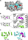
CIB1 ligand binding site and effects of cation binding. A) Surface model of CIB1 showing the hydrophobic pocket, which is thought to be the primary site of protein ligand binding. Hydrophobic pocket residues are indicated by red and cyan. Red-colored residues (Ile-Phe-Asp-Phe117, Leu-Ile153, Phe173) were identified as αIIb-interacting residues (27) and were shown to differentially affect binding to 2 different α-integrin cytoplasmic tails (18). B) Comparison of C-terminal helix (H10) positioning in Ca2+-CIB1, unbound (upper, magenta) and bound to integrin αIIb cytoplasmic tail peptide (lower green). Distances between the α-carbons of residues Leu135 and Ser187 were measured in PyMol. C) Comparison of Mg2+-CIB1 (PDB ID: 2L4I) with Ca2+-CIB1 (PDB ID: 2L4H) showing that the disorderd region from EF-IV through H10 in Mg2+-CIB1 cannot be resolved by NMR spectroscopy. Amino acids are colored blue to green from the N to C terminus, respectively. Divalent cations (either Mg2+ or Ca2+) are colored red in their respective models. At 37°C, αIIb cytoplasmic domain peptide binds to CIB1 with a Kd of 1.41, 1.02, and 3.89 µM in the presence of Ca2+, Mg2+, and neither cation, respectively.

References

    1. Naik U. P., Patel P. M., Parise L. V. (1997) Identification of a novel calcium-binding protein that interacts with the integrin alphaIIb cytoplasmic domain. J. Biol. Chem. 272, 4651–4654 - PubMed
    1. Blamey C. J., Ceccarelli C., Naik U. P., Bahnson B. J. (2005) The crystal structure of calcium- and integrin-binding protein 1: insights into redox regulated functions. Protein Sci. 14, 1214–1221 - PMC - PubMed
    1. Gentry H. R., Singer A. U., Betts L., Yang C., Ferrara J. D., Sondek J., Parise L. V. (2005) Structural and biochemical characterization of CIB1 delineates a new family of EF-hand-containing proteins. J. Biol. Chem. 280, 8407–8415 - PubMed
    1. Huang H., Ishida H., Yamniuk A. P., Vogel H. J. (2011) Solution structures of Ca2+-CIB1 and Mg2+-CIB1 and their interactions with the platelet integrin alphaIIb cytoplasmic domain. J. Biol. Chem. 286, 17181–17192 - PMC - PubMed
    1. Huang H., Vogel H. J. (2012) Structural basis for the activation of platelet integrin αIIbβ3 by calcium- and integrin-binding protein 1. J. Am. Chem. Soc. 134, 3864–3872 - PMC - PubMed

Publication types

MeSH terms

Substances