

Endoscopic Treatment of Early Cancer of the Colon
- PMID: 27118940
- PMCID: PMC4843023
Endoscopic Treatment of Early Cancer of the Colon
Abstract
Colorectal cancer is the fourth most common cancer diagnosis worldwide and the second leading cause of cancer death. In the United States, it is estimated that in 2015 there will be 132,700 new cases of colorectal cancer (representing 8.43% of all new cancer cases) and 49,700 deaths. Colonoscopy plays a fundamental role in the prevention and management of colorectal cancer patients and is used for both the diagnosis and treatment of early colorectal cancer and its precursors. Improvements in colonoscopy preparation, new techniques of adenoma detection, and recent progress in endoscopic imaging methods are providing higher-quality results and reducing the incidence and mortality of the disease. Traditionally, colonoscopy has been used to remove precursor lesions. Invasive cancer was treated by surgical resection with or without chemoradiotherapy. During the past decade, endoscopic resection techniques have advanced, and cancers confined to the mucosal and superficial submucosal layers can now be resected via flexible endoscopes. Therefore, it is important to understand the indications and limitations of endoscopic resection, determine whether the cancer can be curatively resected, and assess the risk of lymph node metastasis, which precludes endoscopic treatment.
Keywords: Colon cancer; adenoma; colonoscopy; endoscopic mucosal resection; endoscopic submucosal dissection; resection.
Figures
Similar articles
-
AGA Institute Clinical Practice Update: Endoscopic Submucosal Dissection in the United States.Clin Gastroenterol Hepatol. 2019 Jan;17(1):16-25.e1. doi: 10.1016/j.cgh.2018.07.041. Epub 2018 Aug 2. Clin Gastroenterol Hepatol. 2019. PMID: 30077787 Review.
-
Clinicopathological features, diagnosis, and treatment of sessile serrated adenoma/polyp with dysplasia/carcinoma.J Gastroenterol Hepatol. 2019 Oct;34(10):1685-1695. doi: 10.1111/jgh.14752. Epub 2019 Jun 30. J Gastroenterol Hepatol. 2019. PMID: 31158302 Review.
-
Colorectal endoscopic submucosal dissection: Technical advantages compared to endoscopic mucosal resection and minimally invasive surgery.Dig Endosc. 2014 Jan;26 Suppl 1:52-61. doi: 10.1111/den.12196. Epub 2013 Nov 5. Dig Endosc. 2014. PMID: 24191896 Review.
-
Early Outcomes of Endoscopic Submucosal Dissection for Colorectal Neoplasms According to Clinical Indications.Dis Colon Rectum. 2016 May;59(5):403-10. doi: 10.1097/DCR.0000000000000549. Dis Colon Rectum. 2016. PMID: 27050602 Clinical Trial.
-
[Endoscopic prevention of colorectal cancer - detection, therapy and control intervals].Ther Umsch. 2018;75(10):583-591. doi: 10.1024/0040-5930/a001044. Ther Umsch. 2018. PMID: 31232661 German.
Cited by
-
Endoscopic non-ablative fractional laser therapy in an orthotopic colon tumour model.Sci Rep. 2018 Jan 26;8(1):1673. doi: 10.1038/s41598-018-19792-2. Sci Rep. 2018. PMID: 29374265 Free PMC article.
-
New plasma diagnostic markers for colorectal cancer: transporter fragments of glutamate tRNA origin.J Cancer. 2024 Jan 12;15(5):1299-1313. doi: 10.7150/jca.92102. eCollection 2024. J Cancer. 2024. PMID: 38356701 Free PMC article.
-
Endoscopic resection of superficial bowel neoplasia: The unmet needs in the Egyptian practice.World J Gastrointest Endosc. 2022 Apr 16;14(4):235-249. doi: 10.4253/wjge.v14.i4.235. World J Gastrointest Endosc. 2022. PMID: 35634486 Free PMC article.
-
Using of endoscopic polypectomy in patients with diagnosed malignant colorectal polyp - The cross-sectional clinical study.Open Med (Wars). 2023 Oct 17;18(1):20230811. doi: 10.1515/med-2023-0811. eCollection 2023. Open Med (Wars). 2023. PMID: 37873541 Free PMC article.
-
A task and performance analysis of endoscopic submucosal dissection (ESD) surgery.Surg Endosc. 2019 Feb;33(2):592-606. doi: 10.1007/s00464-018-6379-6. Epub 2018 Aug 20. Surg Endosc. 2019. PMID: 30128824 Free PMC article.
References
-
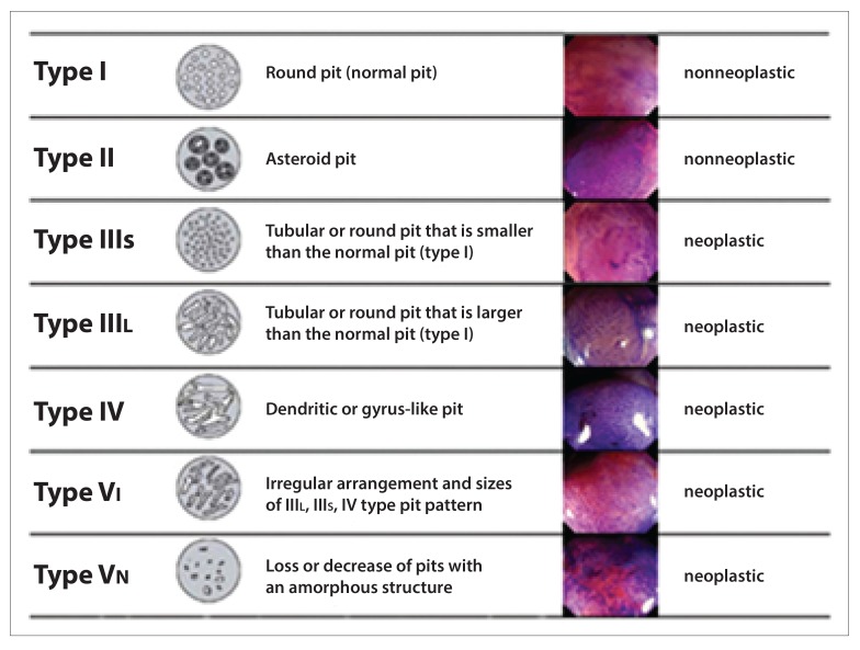
- Tanaka S, Oka S, Kaneko I, et al. Endoscopic submucosal dissection for colorectal neoplasia: possibility of standardization. Gastrointest Endosc. 2007;66(1):100–107. - PubMed
-
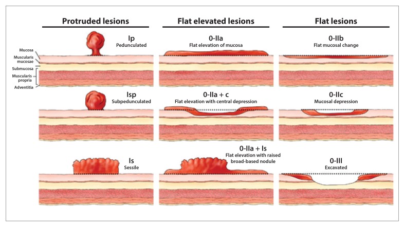
- Endoscopic Classification Review Group. Update on the Paris classification of superficial neoplastic lesions in the digestive tract. Endoscopy. 2005;37(6):570–578. - PubMed
-
- Puli SR, Bechtold ML, Reddy JBK, Choudhary A, Antillon MR. Can endoscopic ultrasound predict early rectal cancers that can be resected endoscopically? A meta-analysis and systematic review. Dig Dis Sci. 2010;55(5):1221–1229. - PubMed
-
- The Paris endoscopic classification of superficial neoplastic lesions: esophagus, stomach, and colon: November 30 to December 1, 2002. Gastrointest Endosc. 2003;58(6 suppl):S3–S43. - PubMed
LinkOut - more resources
Full Text Sources
Miscellaneous