. 2016 May;36(5):302-16.
doi: 10.1089/jir.2015.0091. Epub 2016 Jan 27.

IL-27 Promotes Proliferation of Human Leukemic Cell Lines Through the MAPK/ERK Signaling Pathway and Suppresses Sensitivity to Chemotherapeutic Drugs

Affiliations

IL-27 Promotes Proliferation of Human Leukemic Cell Lines Through the MAPK/ERK Signaling Pathway and Suppresses Sensitivity to Chemotherapeutic Drugs

Haiyan Jia et al. J Interferon Cytokine Res. 2016 May.

Abstract

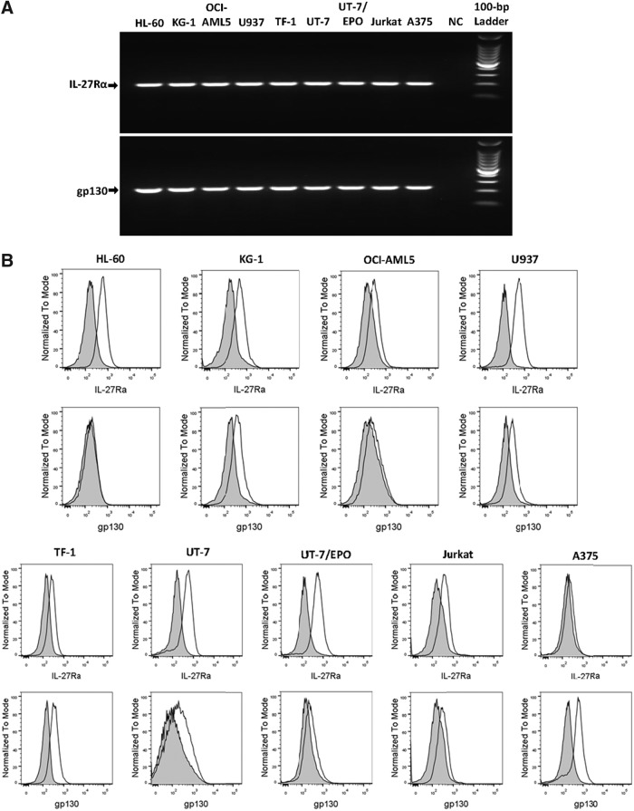
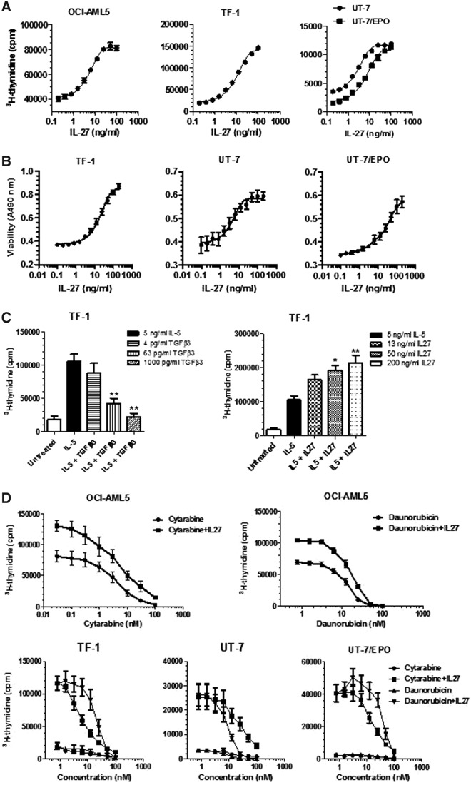
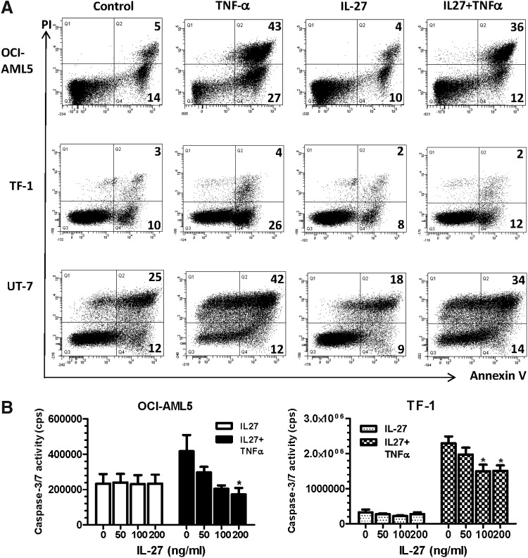
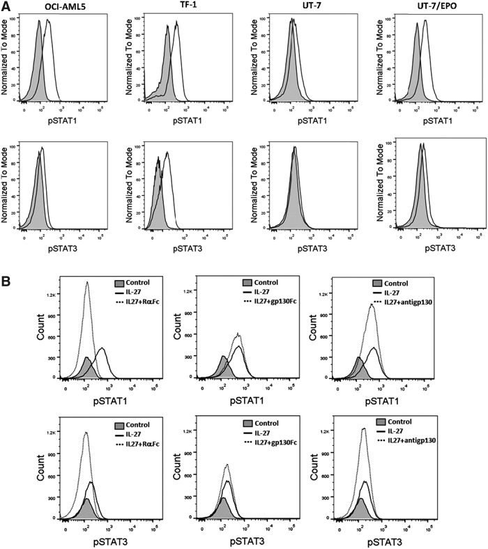
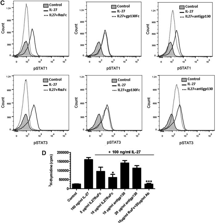
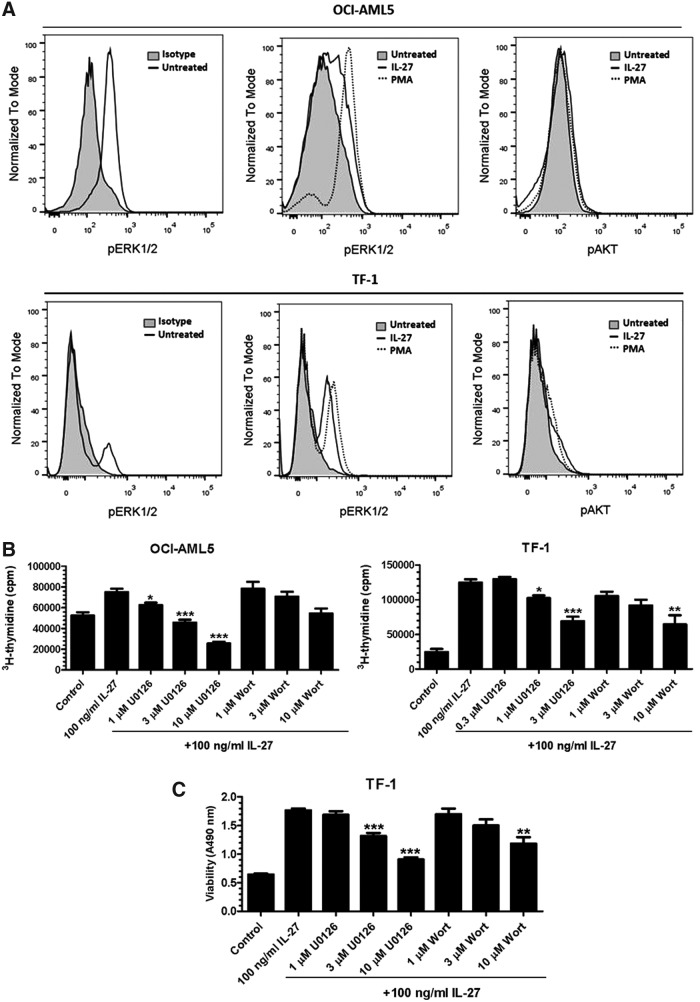
IL-27 is a pleiotropic cytokine of the IL-6/IL-12 family with diverse biological functions. Previous in vivo studies have suggested the antitumor activities of IL-27 in animal models, whereas clinical observations indicate the link of IL-27 in tumor progression. IL-27 has recently been shown to cause inhibition of proliferation on primary leukemic cells from pediatric patients, but information on its role in human leukemic cell lines is limited. In the present study, we investigated the ability of IL-27 to regulate cell growth and survival of various human leukemic cell lines. Our results showed that in human leukemic cell lines coexpressing both IL-27R chains, IL-27Rα and gp130, IL-27 did not inhibit cell growth, but caused dose-dependent proliferation of the acute myeloid leukemic cell line, OCI-AML5, and the erythroleukemic cell lines, TF-1, UT-7, and UT-7/EPO. Consistent with this, IL-27 promoted cell survival and reduced TNF-α-induced apoptosis of the leukemic cell lines. IL-27 also decreased the responsiveness of the leukemic cells to chemotherapeutic drugs, cytarabine and daunorubicin. We observed that IL-27 induced the activation of STAT1/3 and ERK1/2 in the leukemic cells. Growth stimulation by IL-27 was suppressed by the specific MEK inhibitor, U0126, indicating that IL-27-induced cell proliferation is mainly mediated through the activation of the MAPK/ERK signaling pathway. The present study is the first demonstration of the proliferative and antichemotherapeutic properties of IL-27 in human leukemic cell lines, suggesting that IL-27 can play an unfavorable role in tumor growth and can be an important determinant in the chemoresponsiveness of certain subtypes of human leukemia.


Figures

<b>FIG. 1.</b>
FIG. 1.
Expression of the IL-27R complex on human tumor cell lines. (A) mRNA expression of IL-27Rα and gp130 in 9 human tumor cell lines as indicated was analyzed by RT-PCR using specific primers. NC represents a negative control in which cDNA was replaced by H2O. The PCR products were separated by 2% agarose gel electrophoresis and the molecular weights were determined using the 100-bp DNA ladder. (B) Cell surface expression of IL-27Rα and gp130 on 9 human tumor cell lines as indicated was detected by flow cytometry using PE-conjugated specific antibodies. Shaded histograms are isotype-matched mAb staining. Unshaded histograms are IL-27Rα and gp130 staining, respectively. RT-PCR, reverse transcription polymerase chain reaction.
<b>FIG. 2.</b>
FIG. 2.
Proliferation stimulation and desensitization of human leukemic cells to the chemotherapeutic agents by IL-27. (A) Human leukemic cell lines were deprived of GM-CSF or EPO and cultured in 5% or 10% serum assay medium containing IL-27 at increasing concentrations. Dose–response relationships of IL-27 stimulation to cell proliferation were measured by 3H-thymidine incorporation after 2- or 3-day incubation. (B) Human leukemic cell lines were deprived of GM-CSF or EPO and cultured in 2% serum assay medium containing IL-27 at increasing concentrations. Dose–response relationships of IL-27 treatment to cell viability were measured by formazan production after 48 h of incubation. (C) TF-1 cells were deprived of GM-CSF and cultured in 5% serum assay medium containing IL-5 in the absence or presence of either TGF-β3 or IL-27 at indicated concentrations. Cell proliferation was measured by 3H-thymidine incorporation after 2-day incubation. **P < 0.01 for IL-5 plus TGF-β3 versus IL-5 alone. *P < 0.05 and **P < 0.01 for IL-5 plus IL-27 versus IL-5 alone. (D) OCI-AML5, TF-1, UT-7, and UT-7/EPO cells were deprived of GM-CSF or EPO and cultured in 5% or 10% serum assay medium containing cytarabine or daunorubicin at the indicated concentrations in the absence or presence of 200 ng/mL IL-27. Cell proliferation was measured by 3H-thymidine incorporation after 2-day incubation.
<b>FIG. 3.</b>
FIG. 3.
Inhibition of TNF-α-induced apoptosis by IL-27. (A) Leukemic cell lines were deprived of GM-CSF and cultured in serum-free assay medium containing 0.1% BSA in the absence or presence of 10 ng/mL TNF-α, 200 ng/mL IL-27, or 10 ng/mL TNF-α together with 200 ng/mL IL-27 for 4 h and 24 h. Following dual-staining with Annexin V-FITC and PI, percentages of apoptotic cells were determined by flow cytometric analysis of Annexin V+ cells. The Annexin V+ cells at early and late stages of apoptosis are shown as percentages in the lower right quadrant (Q4:% Annexin V+/PI−) and the upper right quadrant (Q2:% Annexin V+/PI+), respectively. Annexin V-FITC versus PI-PE plots with quadrant gates shown are representative of 3 independent experiments yielding very similar results. (B) OCI-AML5 and TF-1 cells were deprived of GM-CSF and cultured in serum-free assay medium containing 0.1% BSA in the absence or presence of 5 ng/mL TNF-α, IL-27 at indicated concentrations, or 5 ng/mL TNF-α together with IL-27 for 2.5 h. At the end of the treatment, the cells were lysed in Caspase-Glo 3/7 substrate and caspase-3/7 activities were assessed by the Caspase-Glo assay. *P < 0.05 for TNF-α plus IL-27 versus TNF-α alone.
<b>FIG. 4.</b>
FIG. 4.
IL-27-activated STAT pathway was mediated through the receptor complex. (A) Intracellular phosphorylation of STAT1 and STAT3 activated by IL-27 was detected by flow cytometry on leukemic cell lines as indicated. Shaded histograms are untreated controls. Unshaded histograms are STAT1-pTyr701 (top) and STAT3-pTyr705 (bottom) staining, respectively. (B, C) IL-27 was preincubated for 15 min with either IL-27Rα-Fc or gp130-Fc before addition to OCI-AML5 (B) and TF-1 (C) cells for 15-min treatment. OCI-AML5 (B) and TF-1 (C) cells were pretreated with anti-gp130 Ab for 15 min, followed by IL-27 stimulation. Intracellular phosphorylation of STAT1 and STAT3 was analyzed by flow cytometry. (D) TF-1 cells were deprived of GM-CSF in 5% serum assay medium containing IL-27 in the absence or presence of either IL-27Rα-Fc or anti-gp130 Ab at indicated concentrations. Cell proliferation was measured by 3H-thymidine incorporation after 2-day incubation. *P < 0.05 for IL-27 plus IL-27Rα-Fc versus IL-27 alone. ***P < 0.001 for IL-27 plus both IL-27Rα-Fc and anti-gp130 Ab versus IL-27 alone.
<b>FIG. 5.</b>
FIG. 5.
Kinetic binding interactions of IL-27 with each receptor chain and of anti-gp130 Ab with gp130. SPR sensorgrams were obtained from injections of IL-27 at concentrations of 0.25, 0.5, 1, 2, 4, and 8 nM over IL-27Rα-Fc captured surface (A), human IL-27 at concentrations of 2, 4, 8, 16, and 32 nM over gp130-Fc captured surface (B), and of anti-gp130 antibody at concentrations of 6.25, 12.5, 25, 50, 100, and 200 nM over gp130-Fc captured surface (C). Experimental data were globally fitted with a 1:1 Langmuir binding model for the determination of kinetic constants. Each sensorgram shown is representative of 3 independent experiments. Color images available online at www.liebertpub.com/jir
<b>FIG. 6.</b>
FIG. 6.
Activation of the MAPK/ERK and PI3K/AKT pathways is required for IL-27-mediated proliferation and survival. (A) Intracellular phosphorylation of ERK1/2-Thr202/Tyr204 and AKT-Ser47 by IL-27 was analyzed by flow cytometry on leukemic cell lines as indicated. (B) OCI-AML5 and TF-1 cells were deprived of GM-CSF and cultured in 10% or 5% serum assay medium containing IL-27 in the absence or presence of the MAPK/ERK inhibitor, U0126, or the PI3K/AKT inhibitor, wortmannin (Wort), at indicated concentrations. Cell proliferation was measured by 3H-thymidine incorporation after 2-day incubation. *P < 0.05, **P < 0.01 and ***P < 0.001 for IL-27 plus U0126 or Wort versus IL-27 alone. (C) TF-1 cells were deprived of GM-CSF and cultured in 2% serum assay medium containing IL-27 in the absence or presence of the MAPK/ERK inhibitor, U0126, or the PI3K/AKT inhibitor, wortmannin (Wort), at indicated concentrations. Cell viability was measured by formazan production after 48 h of incubation. **P < 0.01 and ***P < 0.001 for IL-27 plus U0126 or Wort versus IL-27 alone.

References

    1. Adamopoulos IE, Pflanz S. 2013. The emerging role of Interleukin 27 in inflammatory arthritis and bone destruction. Cytokine Growth Factor Rev 24:115–121 - PMC - PubMed
    1. Awasthi A, Carrier Y, Peron JP, Bettelli E, Kamanaka M, Flavell RA, Kuchroo VK, Oukka M, Weiner HL. 2007. A dominant function for interleukin 27 in generating interleukin 10-producing anti-inflammatory T cells. Nat Immunol 8:1380–1389 - PubMed
    1. Canale S, Cocco C, Frasson C, Seganfreddo E, Di Carlo E, Ognio E, Sorrentino C, Ribatti D, Zorzoli A, Basso G, Dufour C, Airoldi I. 2011. Interleukin-27 inhibits pediatric B-acute lymphoblastic leukemia cell spreading in a preclinical model. Leukemia 25:1815–1824 - PubMed
    1. Creutzig U, van den Heuvel-Eibrink MM, Gibson B, Dworzak MN, Adachi S, de Bont E, Harbott J, Hasle H, Johnston D, Kinoshita A, Lehrnbecher T, Leverger G, Mejstrikova E, Meshinchi S, Pession A, Raimondi SC, Sung L, Stary J, Zwaan CM, Kaspers GJ, Reinhardt D. 2012. AML Committee of the International BFM Study Group. Diagnosis and management of acute myeloid leukemia in children and adolescents: recommendations from an international expert panel. Blood 120:3187–3205 - PubMed
    1. Diakowska D, Lewandowski A, Markocka-Mączka K, GrAbowski K. 2013. Concentration of serum interleukin-27 increase in patients with lymph node metastatic gastroesophageal cancer. Adv Clin Exp Med 22:683–691 - PubMed

MeSH terms

Substances

LinkOut - more resources