Periprosthetic tibial bone mineral density changes after total knee arthroplasty
- PMID: 27120266
- PMCID: PMC4900090
- DOI: 10.3109/17453674.2016.1173982
Periprosthetic tibial bone mineral density changes after total knee arthroplasty
Erratum in
-
Corrigendum.Acta Orthop. 2016 Aug;87(4):x. doi: 10.1080/17453674.2016.1189150. Epub 2016 May 16. Acta Orthop. 2016. PMID: 27181435 Free PMC article. No abstract available.
Abstract
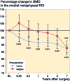
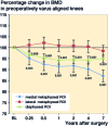
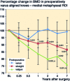
Background and purpose - Total knee arthroplasty (TKA) may cause postoperative periprosthetic bone loss due to stress shielding. Bone also adapts to mechanical alterations such as correction of malalignment. We investigated medium-term changes in bone mineral density (BMD) in tibial periprosthetic bone after TKA. Patients and methods - 86 TKA patients were prospectively measured with dual-energy X-ray absorptiometry (DXA), the baseline measurement being within 1 week after TKA and the follow-up measurements being at 3 and 6 months, and at 1, 2, 4, and 7 years postoperatively. Long standing radiographs were taken and clinical evaluation was done with the American Knee Society (AKS) score. Results - The baseline BMD of the medial tibial metaphyseal region of interest (ROI) was higher in the varus aligned knees (25%; p < 0.001). Medial metaphyseal BMD decreased in subjects with preoperatively varus aligned knees (13%, p < 0.001) and in those with preoperatively valgus aligned knees (12%, p = 0.02) between the baseline and 7-year measurements. No statistically significant changes in BMD were detected in lateral metaphyseal ROIs. No implant failures or revision surgery due to tibial problems occurred. Interpretation - Tibial metaphyseal periprosthetic bone is remodeled after TKA due to mechanical axis correction, resulting in more balanced bone stock below the tibial tray. The diaphyseal BMD remains unchanged after the initial drop, within 3-6 months. This remodeling process was related to good component survival, as there were no implant failures or revision operations due to tibial problems in this medium-term follow-up.
Figures


Comment on
-
Remodeling of the tibial plateau after knee replacement. CT bone densitometry.Acta Orthop Scand. 1988 Oct;59(5):567-73. doi: 10.3109/17453678809148787. Acta Orthop Scand. 1988. PMID: 3188864
-
Long-term changes in bone mineral density following total knee replacement.Clin Orthop Relat Res. 1995 Dec;(321):68-72. Clin Orthop Relat Res. 1995. PMID: 7497687 Clinical Trial.
-
Changes in bone mineral density of the proximal tibia after uncemented total knee arthroplasty. A 3-year follow-up of 25 knees.Acta Orthop Scand. 1995 Dec;66(6):513-6. doi: 10.3109/17453679509002305. Acta Orthop Scand. 1995. PMID: 8553818
-
Bone assessment after total knee arthroplasty by dual-energy X-ray absorptiometry: analysis protocol and reproducibility.Calcif Tissue Int. 1998 Apr;62(4):359-61. doi: 10.1007/s002239900444. Calcif Tissue Int. 1998. PMID: 9504962
-
Finite element analysis of the implanted proximal tibia: a relationship between the initial cancellous bone stresses and implant migration.J Biomech. 1998 Apr;31(4):303-10. doi: 10.1016/s0021-9290(98)00022-0. J Biomech. 1998. PMID: 9672083
-
Dynamic knee loads during gait predict proximal tibial bone distribution.J Biomech. 1998 May;31(5):423-30. doi: 10.1016/s0021-9290(98)00028-1. J Biomech. 1998. PMID: 9727339
-
Bone mineral and migratory patterns in uncemented total knee arthroplasties: a randomized 5-year follow-up study of 38 knees.Acta Orthop Scand. 1999 Dec;70(6):603-8. doi: 10.3109/17453679908997850. Acta Orthop Scand. 1999. PMID: 10665727 Clinical Trial.
-
Measurement of bone density around total knee arthroplasty using fan-beam dual energy X-ray absorptiometry.Calcif Tissue Int. 2000 Sep;67(3):267-72. doi: 10.1007/s002230001111. Calcif Tissue Int. 2000. PMID: 10954783
-
Changes in bone density after cemented total knee arthroplasty: influence of stem design.J Arthroplasty. 2001 Jan;16(1):107-11. doi: 10.1054/arth.2001.16486. J Arthroplasty. 2001. PMID: 11172279
-
Relationships among bone mineral densities, static alignment and dynamic load in patients with medial compartment knee osteoarthritis.Rheumatology (Oxford). 2001 May;40(5):499-505. doi: 10.1093/rheumatology/40.5.499. Rheumatology (Oxford). 2001. PMID: 11371657
-
Increased knee joint loads during walking are present in subjects with knee osteoarthritis.Osteoarthritis Cartilage. 2002 Jul;10(7):573-9. doi: 10.1053/joca.2002.0797. Osteoarthritis Cartilage. 2002. PMID: 12127838
-
Bone morphology in relation to the migration of porous-coated anatomic knee arthroplasties : a roentgen stereophotogrammetric and histomorphometric study in 23 knees.J Arthroplasty. 2003 Aug;18(5):649-53. doi: 10.1016/s0883-5403(03)00111-6. J Arthroplasty. 2003. PMID: 12934220 Clinical Trial.
-
Periprosthetic tibial bone mineral density changes after total knee arthroplasty: one-year follow-up study of 69 patients.Acta Orthop Scand. 2004 Oct;75(5):600-5. doi: 10.1080/00016470410001493. Acta Orthop Scand. 2004. PMID: 15513494
-
Effect of hydroxyapatite-coated tibial components on changes in bone mineral density of the proximal tibia after uncemented total knee arthroplasty: a prospective randomized study using dual-energy x-ray absorptiometry.J Arthroplasty. 2005 Jun;20(4):516-20. doi: 10.1016/j.arth.2004.09.041. J Arthroplasty. 2005. PMID: 16124970 Clinical Trial.
-
Peri-prosthetic bone mineral density after total knee arthroplasty. Cemented versus cementless fixation.J Bone Joint Surg Br. 2006 May;88(5):606-13. doi: 10.1302/0301-620X.88B5.16893. J Bone Joint Surg Br. 2006. PMID: 16645105
-
Aseptic loosening, not only a question of wear: a review of different theories.Acta Orthop. 2006 Apr;77(2):177-97. doi: 10.1080/17453670610045902. Acta Orthop. 2006. PMID: 16752278 Review.
-
Contribution of loading conditions and material properties to stress shielding near the tibial component of total knee replacements.J Biomech. 2007;40(6):1410-6. doi: 10.1016/j.jbiomech.2006.05.020. Epub 2006 Jul 17. J Biomech. 2007. PMID: 16846605
-
Influence of the tibial stem design on bone density after cemented total knee arthroplasty: a prospective seven-year follow-up study.Int Orthop. 2008 Feb;32(1):47-51. doi: 10.1007/s00264-006-0280-y. Epub 2006 Nov 18. Int Orthop. 2008. PMID: 17115154 Free PMC article.
-
Joint area constraint had no influence on bone loss in proximal tibia 5 years after total knee replacement.J Orthop Res. 2007 Jun;25(6):798-803. doi: 10.1002/jor.20358. J Orthop Res. 2007. PMID: 17318893
-
Medial-to-lateral ratio of tibiofemoral subchondral bone area is adapted to alignment and mechanical load.Calcif Tissue Int. 2009 Mar;84(3):186-94. doi: 10.1007/s00223-008-9208-4. Epub 2009 Jan 16. Calcif Tissue Int. 2009. PMID: 19148562 Free PMC article.
-
Loss of tibial bone density in patients with rotating- or fixed-platform TKA.Clin Orthop Relat Res. 2010 Mar;468(3):775-81. doi: 10.1007/s11999-009-0794-x. Epub 2009 Mar 26. Clin Orthop Relat Res. 2010. PMID: 19322618 Free PMC article. Clinical Trial.
References
-
- Abu-Rajab R B, Watson W S, Walker B, Roberts J, Gallacher S J, Meek R M.. Peri-prosthetic bone mineral density after total knee arthroplasty. cemented versus cementless fixation. J Bone Joint Surg Br 2006; 88 (5): 606–13. - PubMed
-
- Au A G, James Raso V, Liggins A B, Amirfazli A.. Contribution of loading conditions and material properties to stress shielding near the tibial component of total knee replacements. J Biomech 2007; 40 (6): 1410–6. - PubMed
-
- Baliunas A J, Hurwitz D E, Ryals A B, Karrar A, Case J P, Block J A, Andriacchi T P.. Increased knee joint loads during walking are present in subjects with knee osteoarthritis. Osteoarthritis Cartilage 2002; 10 (7): 573–9. - PubMed
-
- Bohr H H, Schaadt O.. Mineral content of upper tibia assessed by dual photon densitometry. Acta Orthop Scand 1987; 58 (5): 557–9. - PubMed
Publication types
MeSH terms
LinkOut - more resources
Full Text Sources
Other Literature Sources
Medical
