Review
. 2016 Jun;46(6):1315-25.
doi: 10.1002/eji.201545562. Epub 2016 May 17.

Regulation of metabolic health and adipose tissue function by group 2 innate lymphoid cells

Affiliations
Review

Regulation of metabolic health and adipose tissue function by group 2 innate lymphoid cells

Kelly M Cautivo et al. Eur J Immunol. 2016 Jun.

Abstract

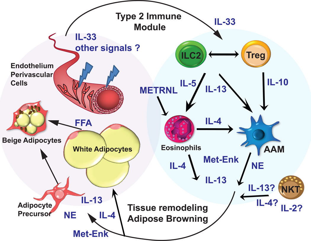
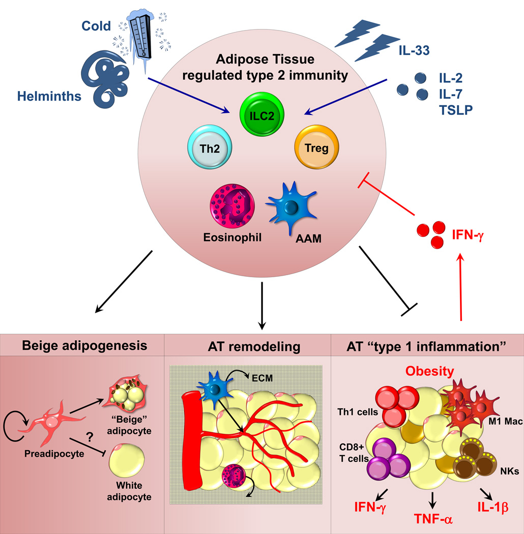
Adipose tissue (AT) is home to an abundance of immune cells. With chronic obesity, inflammatory immune cells accumulate and promote insulin resistance and the progression to type 2 diabetes mellitus. In contrast, recent studies have highlighted the regulation and function of immune cells in lean, healthy AT, including those associated with type 2 or "allergic" immunity. Although traditionally activated by infection with multicellular helminthes, AT type 2 immunity is active independently of infection, and promotes tissue homeostasis, AT "browning," and systemic insulin sensitivity, protecting against obesity-induced metabolic dysfunction and type 2 diabetes mellitus. In particular, group 2 innate lymphoid cells (ILC2s) are integral regulators of AT type 2 immunity, producing the cytokines interleukin-5 and IL-13, promoting eosinophils and alternatively activated macrophages, and cooperating with and promoting AT regulatory T (Treg) cells. In this review, we focus on the recent developments in our understanding of group 2 innate lymphoid cell cells and type 2 immunity in AT metabolism and homeostasis.

Keywords: Adipose tissue; Diabetes; Group 2 innate lymphoid cells; Metabolism; Type 2 immunity.


Conflict of interest statement

The authors declare no financial or commercial conflict of interest.

Figures

Figure 1
Figure 1. Model of the interaction of type 2 immunity and healthy adipose tissue
Adipose tissue ILC2s produce IL-5 and IL-13, whereas Treg cells produce IL-10, and together these cells and cytokines coordinate recruitment and maintenance of eosinophils and AAMs right). ILC2s and NKT cells are required for Treg-cell maintenance in the AT, and NKT cells may be an additional source of IL-4 and IL-13. Eosinophis can produce IL-4 and are required for optimal AAM maintenance. Together, this “regulated” type 2 immune response (right) acts coordinately on the adipose tissue to promote browning and other adipose tissue remodeling (left). IL-4 and/or IL-13 promote adipocyte precursors to replicate and undergo beige differentiation. Met-Enk and AAM-derived norepinephrine (NE) further support beige adipocytes, as well as free fatty acid release from white adipocytes. In turn, adipose endothelium and supporting cells release IL-33, and other unknown chemokines and cytokines, that support the numbers and function of ILC2s and Treg cells. ILC2: group 2 innate lymphoid cells; Treg: regulatory T cell; AAM: alternatively activated macrophage; NE: norepinephrine; FFA: free fatty acids; Met-Enk: methionine encephalin.
Figure 2
Figure 2. Model of regulators of adipose tissue homeostatic immune cells
Positive signals enhancing numbers or function of “regulated” type 2 immunity within the AT are depicted in blue. IL-33 directly activates and supports ILC2 and Treg cells. Helminths and cold enhance responses through currently unknown mechanisms. IL-2, IL-7, and TSLP are speculated to be positive signals in AT. Signals that restrict this AT immune module are noted in red. Obesity-associated IFN-γ directly represses ILC2 and possibly other cells noted, including Treg cells. On bottom, three possible mechanisms by which type 2 immunity could impact AT function are illustrated: 1) promoting AT browning 2) promoting AT physiologic remodeling or 3) restricting excess or maladaptive “type 1 inflammation”. NK: nature killer cells; M1 Mac: M1 macrophage; ECM: extracellular matrix.

References

    1. Ng M, Fleming T, Robinson M, Thomson B, Graetz N, Margono C, Mullany EC, et al. Global, regional, and national prevalence of overweight and obesity in children and adults during 1980–2013: a systematic analysis for the Global Burden of Disease Study 2013. Lancet. 2014;384:766–781. - PMC - PubMed
    1. Ouchi N, Parker JL, Lugus JJ, Walsh K. Adipokines in inflammation and metabolic disease. Nat Rev Immunol. 2011;11:85–97. - PMC - PubMed
    1. Wensveen FM, Valentic S, Sestan M, Turk Wensveen T, Polic B. The "Big Bang" in obese fat: Events initiating obesity-induced adipose tissue inflammation. Eur J Immunol. 2015;45:2446–2456. - PubMed
    1. Bapat SP, Myoung Suh J, Fang S, Liu S, Zhang Y, Cheng A, Zhou C, et al. Depletion of fat-resident Treg cells prevents age-associated insulin resistance. Nature. 2015;528:137–141. - PMC - PubMed
    1. Asterholm IW, Tao C, Morley TS, Wang QA, Delgado-Lopez F, Wang ZV, Scherer PE. Adipocyte inflammation is essential for healthy adipose tissue expansion and remodeling. Cell Metabolism. 2014:103–118. - PMC - PubMed

Publication types