. 2016 Apr 28:6:25146.
doi: 10.1038/srep25146.

Generation of siRNA Nanosheets for Efficient RNA Interference

Affiliations

Generation of siRNA Nanosheets for Efficient RNA Interference

Hyejin Kim et al. Sci Rep. .

Abstract

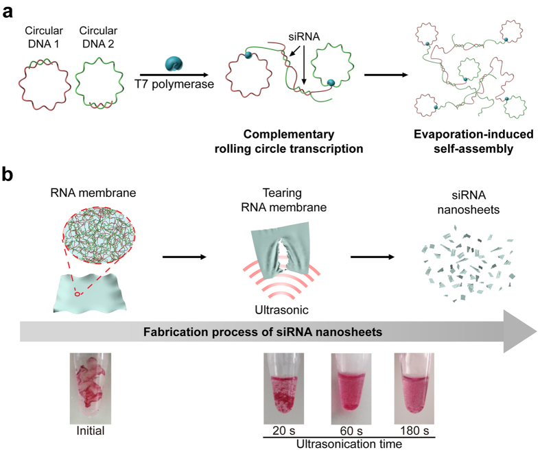
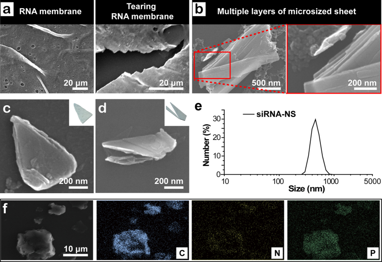
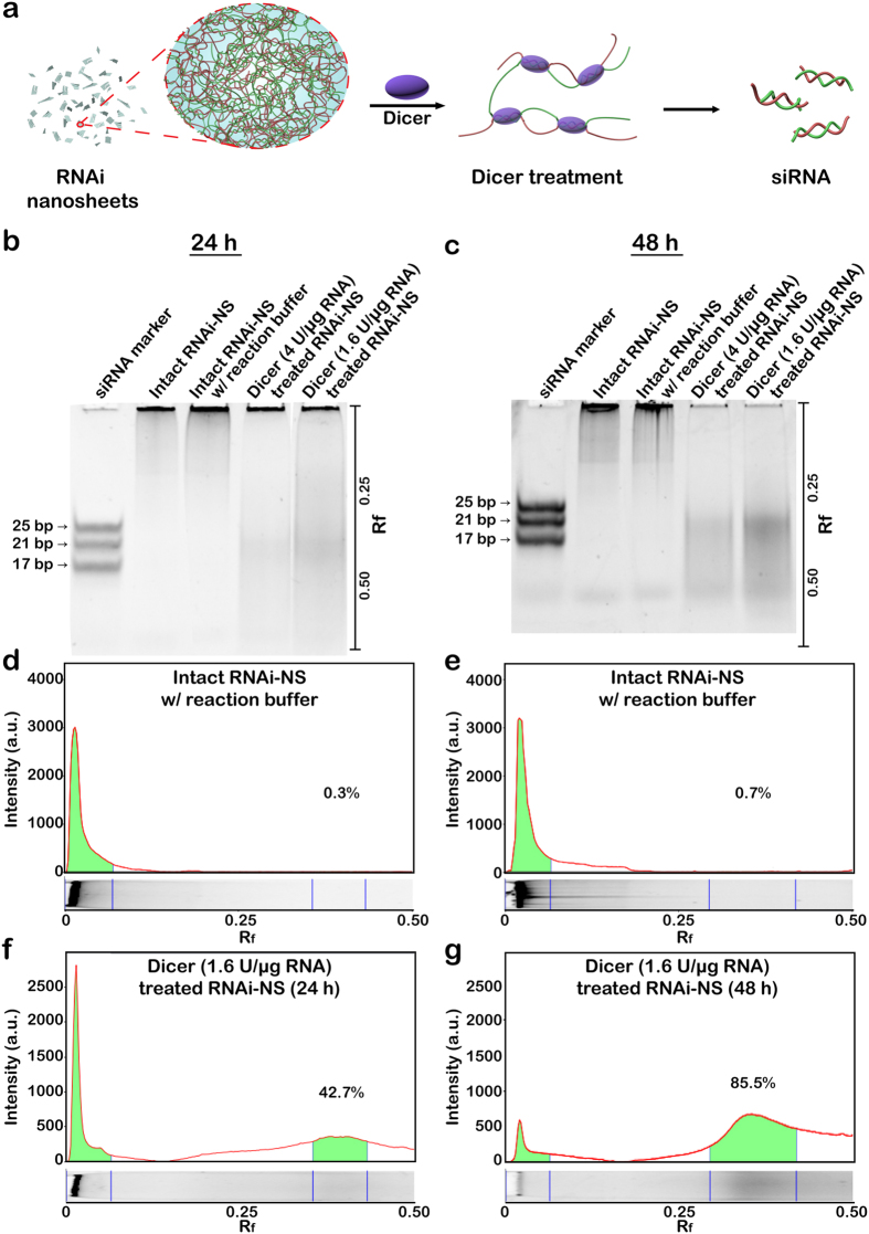
After the discovery of small interference RNA (siRNA), nanostructured siRNA delivery systems have been introduced to achieve an efficient regulation of the target gene expression. Here we report a new siRNA-generating two dimensional nanostructure in a formation of nanosized sheet. Inspired by tunable mechanical and functional properties of the previously reported RNA membrane, siRNA nanosized sheets (siRNA-NS) with multiple Dicer cleavage sites were prepared. The siRNA-NS has two dimensional structure, providing a large surface area for Dicer to cleave the siRNA-NS for the generation of functional siRNAs. Furthermore, downregulation of the cellular target gene expression was achieved by delivery of siRNA-NS without chemical modification of RNA strands or conjugation to other substances.


Figures

Figure 1
Figure 1. Schematic illustration of the synthetic process of siRNA-NS.
(a) Design for siRNA-NS bearing siRNA precursors. Complementary rolling circle transcription (cRCT) is carried out by T7 RNA polymerase with circular DNA1 and circular DNA2, partially complementary to circular DNA1. The cRCT process is followed by evaporation-induced self-assembly, completing the synthetic process of RNA membrane. (b) Schematic illustration and digital images of GelRed-stained RNA membrane after ultrasonication. As sonication proceeds, RNA membrane lost its initial form and torn into large pieces (~20 s). After 60 s of sonication, RNA membrane was broken into smaller pieces with large sediments still remaining. At the time point of 180 s after sonication, no sediment was observed with naked eye.
Figure 2
Figure 2. Characterization of the siRNA-NS.
(a) SEM images of the surface and the site being torn of RNA membrane. (b) SEM images of the microsized RNA sheet after 10 min of sonication, and high magnification image of the highlighted area, revealing its multi-layered structure. (c–d) High magnification SEM images of siRNA-NS and schematic illustrations of each shape of the nanosheets (inset). (e) Size distribution of anti-GFP siRNA-NS obtained with dynamic light scattering analysis. (f ) SEM-based EDS mapping of siRNA-NS, revealing its composition.
Figure 3
Figure 3. Synthesis of siRNA from siRNA-NS.
(a) Schematic illustration of siRNA production from siRNA-NS. (b,c) Gel electrophoresis analysis of siRNA-NS treated with Dicer enzyme for 24 h (b) and 48 h (c). Lanes 1-3 indicate siRNA marker, untreated siRNA-NS (114 ng) and untreated siRNA-NS with Dicer reaction buffer, respectively. Lanes 4-5 indicate the same amount of siRNA-NS treated with 4 units or 1.6 units per μg RNA, respectively. (d–g) Band intensities of the lane 3 (d,e) and lane 5 (f,g) in each gel were analyzed. The values of Rf were indicated for the exact determination of the positions of the bands.
Figure 4
Figure 4. siRNA-NS-mediated gene knockdown assay.
(a) Fluorescence microscopy images of HeLa-GFP cells at 24 h after treatment with 100 pM, 250 pM and 500 pM of anti-GFP siRNA-NS. Control cells were left untreated for 24 h. (b) GFP expressions of HeLa-GFP cells at 24 h after treatment with 20 pM, 100 pM, 250 pM and 500 pM of anti-GFP siRNA-NS (orange), scrambled siRNA-NS (green) or left untreated (black). GFP intensities were normalized with the intensity of untreated cells (n = 4). (c) Image cytometry analysis of anti-GFP siRNA-NS treated HeLa-GFP cells at 12 h (yellow) and 24 h (orange) after the treatment.

References

    1. Breaker R. R. Natural and engineered nucleic acids as tools to explore biology. Nature 432, 838–845 (2004). - PubMed
    1. Grabow W. W. & Jaeger L. RNA self-assembly and RNA nanotechnology. Acc Chem Res 47, 1871–1880 (2014). - PubMed
    1. Guo P., Haque F., Hallahan B., Reif R. & Li H. Uniqueness, advantages, challenges, solutions, and perspectives in therapeutics applying RNA nanotechnology. Nucleic Acid Ther 22, 226–245 (2012). - PMC - PubMed
    1. Kim H. et al.. Nucleic acid engineering: RNA following the trail of DNA. ACS Comb Sci 18, 87–99 (2016). - PubMed
    1. Fire A. et al.. Potent and specific genetic interference by double-stranded RNA in Caenorhabditis elegans. Nature 391, 806–811 (1998). - PubMed

Publication types

MeSH terms

Substances