. 2016 Apr 28:7:11449.
doi: 10.1038/ncomms11449.

An essential malaria protein defines the architecture of blood-stage and transmission-stage parasites

Affiliations

An essential malaria protein defines the architecture of blood-stage and transmission-stage parasites

Sabrina Absalon et al. Nat Commun. .

Abstract

Blood-stage replication of the human malaria parasite Plasmodium falciparum occurs via schizogony, wherein daughter parasites are formed by a specialized cytokinesis known as segmentation. Here we identify a parasite protein, which we name P. falciparum Merozoite Organizing Protein (PfMOP), as essential for cytokinesis of blood-stage parasites. We show that, following PfMOP knockdown, parasites undergo incomplete segmentation resulting in a residual agglomerate of partially divided cells. While organelles develop normally, the structural scaffold of daughter parasites, the inner membrane complex (IMC), fails to form in this agglomerate causing flawed segmentation. In PfMOP-deficient gametocytes, the IMC formation defect causes maturation arrest with aberrant morphology and death. Our results provide insight into the mechanisms of replication and maturation of malaria parasites.


Figures

Figure 1
Figure 1. Generation of PfMOP-DD transgenic parasites.
(a) Schematic of the DD system. PfMOP-DD-fusion protein is targeted for rapid degradation in the absence of Shld1. A haemagglutinin (HA) epitope tag is also present. (b) Protein lysates prepared from ring, trophozoite (Troph) or schizont stage PfMOP-DD parasites cultured with 250 nM Shld1 and probed with antibodies to HA or PfLDH (loading control). (c) Immunoblot of protein lysates from PfMOP-DD schizont stage parasites (36–48 h.p.i.) cultured [+]/[−] 250 nM Shld1 and probed with antibodies to HA, PfRhopH3 (a schizont stage loading control) or PfLDH (loading control). Quantification of immunoblot was performed by volumetric measurement of fluorescence intensity with the LiCor Odyssey CLx system. (d) Replication curves of PfMOP-DD and PfCDPK4-DD parasites cultured [+]/[−] 250 nM Shld1 (n=3, mean with ±s.d. error bars).
Figure 2
Figure 2. PfMOP is required for efficient asexual parasite growth.
Time course of PfMOP-DD parasites cultured [+]/[−] Shld1. Samples were collected every 2 h from 40 to 60 h.p.i. and parasitemia was determined by flow cytometry (n=3, mean with 95% CI error bars).
Figure 3
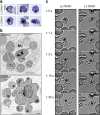
Figure 3. PfMOP is essential for schizont segmentation.
PfMOP-DD parasites were grown [+]/[−] Shld1 until 52 h.p.i. (E64 added for 6 h) and visualized either by light microscopy ((a) scale bar, 1 μm) or by electron microscopy ((b) scale bar, 500 nm). (c). Frames from video microscopy of [+]/[−] Shld1 PfMOP-DD schizont rupture. The first frame with a released merozoite was arbitrarily set at t=1 s. Scale bar, 5 μm. Representative video shown for each condition. Nine rupture events scored for [+] Shld1 with 8 out of 9 having normal egress. Sixteen rupture events scored for [−] Shld1 with 1 out of 16 having normal and 15 out of 16 having aberrant egress.
Figure 4
Figure 4. PfMOP localizes to apical area of parasites.
Representative pictures of E64-treated schizont-stages. (a) Wide-field IFA of segmented schizont stained with antibodies against PfRAMA, PfRON4, and HA. Scale bar, 1 μm. (b) SR-SIM showing deconvolution of single z-section and three-dimensional reconstruction. Scale bar, 1 μm. (c) Transmission electron microscopy with (white arrows) gold-labelled antibodies against HA (N, nucleus; R, rhoptry, scale bar, 100 nm). (d) Proteinase K protection assay. Schizont stage parasites were permeabilized with saponin and [+]/[−] digitonin, [+]/[−] treated with proteinase K and probed with antibodies to HA. Control proteins are luminal rhoptry protein (PfRhopH3), cytoskeleton protein (spectrin) and cytosolic protein (PfLDH).
Figure 5
Figure 5. PfAMA1 translocation is aberrant in schizonts with PfMOP-knockdown.
(a) Schizonts from [+]/[−] Shld1 PfMOP-DD parasites were E64-treated, fixed, probed with anti-PfAMA1 and scored as micronemal (M), partially translocated (PT) or surface (S), representative [−] Shld1 parasite IFAs shown. (b) Chart shows proportions of each type (100 schizonts per condition from n=3 independent experiments; mean with 95% CI error bars; *P<0.01, groups compared by unpaired t-test, scale bar, 1 μm). (c) Synchronized schizont stage (40–44 h) parasites, maintained with 250 nM (left panel) or 0 nM (right panel) Shld1, were incubated 6 h in presence of 10 μM E64, methanol-fixed, permeabilized, and stained using antibodies against PfRON4, PfRhopH3, PfEBA175 and PfTubulin. Staining for these markers was similar in [+] and [−] Shld1 conditions. PfAMA1 staining was used to identify E64-treated schizonts that were sufficiently mature (that is, surface staining or partially translocated staining, but not micronemal staining, scale bar, 1 μm).
Figure 6
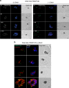
Figure 6. Residual PfMOP in knockdown parasites forms fewer and less bright accumulations.
(a) Synchronized parasites maintained [+] and [−] Shld1 were sampled for processing every 2–4 h from 36 to 44 h.p.i., methanol-fixed, stained with the anti-HA antibody and counterstained with DAPI. Green arrowheads indicate the dimmer foci of PfMOP staining in [−] Shld1 samples. The number of nuclei is shown in upper left corner. (b) Synchronized parasite cultured with Shld1 were fixed and probed with DAPI, anti-HA antibody (green) and anti-GAP45 antibody (red). Images are representative of multiple independent biological replicates. Scale bar, 1 μm.
Figure 7
Figure 7. PfMOP deficiency leads to incomplete formation of the IMC.
(a) Wide-field and (b) SR-SIM of representative pictures of E64-treated [+]/[−] Shld1 schizont stage PfMOP-DD parasites using antibodies against PfGAP45 and PfMSP1. Scale bar, 1 μm.
Figure 8
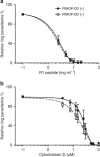
Figure 8. Released merozoites from PfMOP-deficient schizonts invade normally.
Analysis of the sensitivity of PfMOP-DD parasites to inhibition of invasion by either the R1 peptide (a) or cytochalasin D (b). Synchronized schizont stage (42–46 h.p.i.) parasites, maintained [+]/[−] Shld1, were purified, then incubated for an additional 8–12 h with a range of R1 peptide or cytochalasin D concentrations. Newly re-invaded ring-stage parasites were measured by flow cytometry (R1 peptide n=3, cytochalasin D n=2, mean with 95% CI error bars, non-linear fit: log (agonist) versus response EC50).
Figure 9
Figure 9. PfMOP is essential for gametocyte maturation.
(a) Gametocyte conversion rate (n=3; mean with 95% CI error bars, groups compared with unpaired t-test). (b) Prevalence of P. falciparum gametocyte stages in PfMOP1-DD induced and grown [+]/[−] Shld1 by light microscopy (100 gametocytes were counted per condition from n=3 independent experiments; mean with 95% CI error bars; **P<0.001, groups compared with unpaired t-test, scale bar, 2 μm). (c) IFA using antibodies against PfGAP45 and PfTubulin show expected stages/morphology in [+] Shld1 and reveal absence of IMC development and abnormal shape in [−]Shld1 gametocytes, scale bar, 2 μm.
Figure 10
Figure 10. Model of PfMOP function in Plasmodium falciparum asexual life cycle.
The schematic illustrates PfMOP protein localization and function through the asexual and transmission stages of the P. falciparum life cycle. In the left panel, the level of PfMOP protein is maintained by the presence of Shdl1. At the early schizont stage, parasites express PfMOP protein that localizes to the apical area of each newly forming daughter cell. As the schizont matures, PfMOP facilitates formation of the IMC, which leads to complete segmentation of the daughter merozoites through plasma membrane budding. For sexually committed rings, PfMOP expression facilitates IMC formation to maintain the survival and define the shape of the gametocyte. On the right panel, the consequences of PfMOP-deficiency are shown. At the early schizont stage, the quantity and number of PfMOP foci are both decreased. The low-level residual PfMOP is only detectable on a subset of forming daughter parasites, with the remaining trapped as an agglomerate under a common plasma membrane. In the absence of PfMOP, the IMC fails to form in the agglomerate. Egress is triggered normally, but the incompletely segmented merozoites remain attached to the residual body. The gametocyte conversion rate is unchanged by the absence of PfMOP. However, in absence of PfMOP, gametocytes lack an IMC and become aberrantly shaped and ultimately pyknotic.

References

    1. World Health Organization. World Malaria Report 2014, 1–142 (2014).
    1. White N. J. et al.. Malaria. Lancet 383, 723–735 (2014). - PubMed
    1. Francia M. E. & Striepen B. Cell division in apicomplexan parasites. Nat. Rev. Microbiol. 12, 125–136 (2014). - PubMed
    1. Hu K. et al.. Daughter cell assembly in the protozoan parasite Toxoplasma gondii. Mol. Biol. Cell 13, 593–606 (2002). - PMC - PubMed
    1. Tilney L. G. & Tilney M. S. The cytoskeleton of protozoan parasites. Curr. Opin. Cell Biol. 8, 43–48 (1996). - PubMed

Publication types

Substances