. 2016 Jun;13(6):5053-8.
doi: 10.3892/mmr.2016.5191. Epub 2016 Apr 26.

Transplantation of ovarian granulosa‑like cells derived from human induced pluripotent stem cells for the treatment of murine premature ovarian failure

Affiliations

Transplantation of ovarian granulosa‑like cells derived from human induced pluripotent stem cells for the treatment of murine premature ovarian failure

Te Liu et al. Mol Med Rep. 2016 Jun.

Abstract

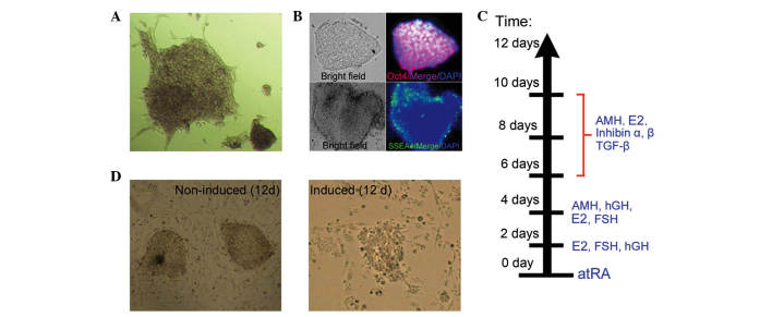
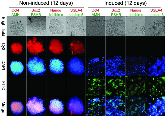
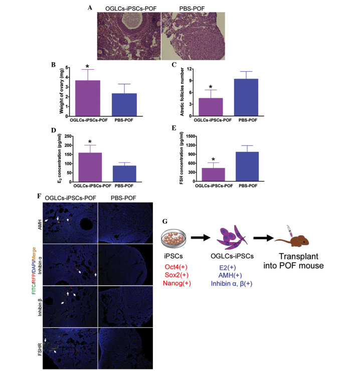
Premature ovarian failure (POF) is a common cause of female infertility, for which there are currently no ideal treatments or medications. Furthermore, apoptosis of ovarian granulosa cells (OGCs) is an important mechanism underlying the decline in ovarian reserve and function. In the present study, several cellular growth factors and hormones were used to induce the differentiation of human induced pluripotent stem cells (iPSCs) into ovarian granulosa‑like cells (OGLCs) in vitro. Immunohistochemical staining demonstrated that OGLCs derived from iPSCs strongly expressed granulosa cell markers, including anti‑Müllerian hormone, inhibin α, inhibin β and follicle‑stimulating hormone receptor, but did not express stem cell markers, including octamer‑binding transcription factor 4, SRY (sex determining region Y)-box 2, Nanog and stage-specific embryonic antigen-4 12 days post‑induction. In addition, a mouse model of POF was generated by cyclophosphamide treatment. Subsequently, iPSC‑derived OGLCs were transplanted into the POF mice (OGLCs‑iPSCs‑POF group) in vivo. Results indicated that, compared with the control group (POF mice treated with phosphate‑buffered saline), the growth state of OGLCs was markedly improved, and mature follicles could be detected in the ovarian tissue of the OGLCs‑iPSCs‑POF group. Immunohistochemical staining demonstrated that iPSC‑derived OGLCs transplanted into POF mice not only exhibited substantial growth in murine ovarian tissues, but also strongly expressed OGC markers. Furthermore, enzyme‑linked immunosorbent assays indicated that the levels of the hormone estradiol in peripheral blood samples were significantly enhanced following transplantation of iPSC‑derived OGLCs into POF mice. Furthermore, ovarian tissue weight was significantly higher in the OGLCs‑iPSCs‑POF group compared with in the control group, and the number of atretic follicles in OGLCs‑iPSCs‑POF mice was significantly reduced, as compared with in the control mice. These results suggest that OGLCs derived from human iPSCs may not only effectively enhance OGC growth and repair damaged ovarian tissue, but may also maintain the ovarian tissue niche, promoting follicular development and maturation in a mouse model of POF.


Figures

Figure 1
Figure 1
'Stemness' of induced pluripotent stem cells (iPSCs) following induction. (A) Alkaline phosphatase (AKP) staining assay indicated that iPSCs exhibited deep blue staining. Original magnification, ×200. (B) Immunofluorescence staining indicated that iPSCs highly expressed octamer-binding transcription factor 4 (Oct4) (red fluorescence) and stage-specific embryonic antigen-4 (SSEA4) (green fluorescence) proteins. Original magnification, ×200. (C) Various cell growth factors and hormones were used at various time points to induce iPSCs to differentiate into ovarian granulosa-like cells. (D) Phenotype of iPSCs during induction. Original magnification, ×200. AMH, anti-Müllerian hormone; E2, estradiol; TGF-β, transforming growth factor-β; hGH, human growth hormone; FSH, follicle-stimulating hormone; atRA, all-trans-retinoic acid.
Figure 2
Figure 2
Immunofluorescence staining. Immunohistochemical staining indicated that ovarian granulosa-like cells derived from induced pluripotent stem cells strongly expressed ovarian granulosa cells markers [anti-Müllerian hormone (AMH), inhibin α, inhibin β and follicle-stimulating hormone receptor (FSHR)], but not stem cell markers [octamer-binding transcription factor 4 (Oct4), SRY (sex-determining region Y)-box 2 (Sox2), Nanog and stage-specific embryonic antigen-4 (SSEA4) 12 days post-induction. Each sample was stained for two markers with the color of the fluorescence indicated by the color of the word. Original magnification, ×200. Cy3, cyanine 3; DAPI, 4′,6-diamidino-2-phenylindole; FITC, fluorescein isothiocyanate.
Figure 3
Figure 3
Effects of induced pluripotent stem cell (iPSC)-derived ovarian granulosa-like cells (OGLCs) transplanted into a mouse model of premature ovarian failure (POF). (A) Hematoxylin-eosin staining and pathological analysis revealed that iPSC-derived OGLCs improved the ovarian tissue niche, reduced the number of atretic follicles, and increased the number of normal follicles. OGLCs-iPSCs-POF, POF mice transplanted with iPSC-induced OGLCs (experimental group); PBS-POF, POF mice treated with PBS (control group). Original magnification, ×200. (B) Ovarian tissue weight was elevated in the OGLCs-iPSCs-POF group compared with in the control group. (C) Number of atretic follicles in the OGLCs-iPSCs-POF group was significantly reduced. (D) Plasma estradiol (E2) levels were determined by enzyme-linked immunosrobent assay (ELISA); E2 levels were higher in the OGLCs-iPSCs-POF group compared with in the PBS-POF group. (E) Plasma follicle-stimulating hormone (FSH) levels were determined by ELISA; FSH levels were lower in the OGLCs-iPSCs-POF group compared with in the PBS-POF group. Data are presented as the mean ± standard error of the mean. *P<0.05 vs. the PBS-POF group; n=15. (F) Immunofluorescence assay indicated that iPSC-derived OGLCs transplanted into POF mice (red fluorescence) expressed OGC biomarkers (green fluorescence) in ovarian tissue. White arrows represent cells with red fluorescence merged with green fluorescence. Original magnification, ×200. (G) iPSCs induction and transplantation process. AMH, anti-Müllerian hormone; Oct4, octamer-binding transcription factor 4; Sox2, SRY (sex-determining region Y)-box 2; FSHR, FSH receptor; FITC, fluorescein isothiocyanate; RFP, red fluorescent protein; DAPI, 4′,6-diamidino-2-phenylindole.

References

    1. Vujović S, Ivović M, Tancić-Gajić M, Marina L, Barać M, Arizanović Z, Nenezić A, Ivanisević M, Micić J, Sajić S, Micić D. Premature ovarian failure. Srp Arh Celok Lek. 2012;140:806–811. doi: 10.2298/SARH1212806V. - DOI - PubMed
    1. Liu T, Huang Y, Guo L, Cheng W, Zou G. CD44+/CD105+ human amniotic fluid mesenchymal stem cells survive and proliferate in the ovary long-term in a mouse model of chemotherapy-induced premature ovarian failure. Int J Med Sci. 2012;9:592–602. doi: 10.7150/ijms.4841. - DOI - PMC - PubMed
    1. Liu T, Qin W, Huang Y, Zhao Y, Wang J. Induction of estrogen-sensitive epithelial cells derived from human-induced pluripotent stem cells to repair ovarian function in a chemotherapy-induced mouse model of premature ovarian failure. DNA Cell Biol. 2013;32:685–698. doi: 10.1089/dna.2013.2032. - DOI - PMC - PubMed
    1. Liu T, Huang Y, Zhang J, Qin W, Chi H, Chen J, Yu Z, Chen C. Transplantation of human menstrual blood stem cells to treat premature ovarian failure in mouse model. Stem Cells Dev. 2014;23:1548–1557. doi: 10.1089/scd.2013.0371. - DOI - PMC - PubMed
    1. Takahashi K, Yamanaka S. Induction of pluripotent stem cells from mouse embryonic and adult fibroblast cultures by defined factors. Cell. 2006;126:663–676. doi: 10.1016/j.cell.2006.07.024. - DOI - PubMed

MeSH terms

Substances