

Partial meniscectomy is associated with increased risk of incident radiographic osteoarthritis and worsening cartilage damage in the following year
- PMID: 27121931
- PMCID: PMC5083232
- DOI: 10.1007/s00330-016-4361-z
Partial meniscectomy is associated with increased risk of incident radiographic osteoarthritis and worsening cartilage damage in the following year
Abstract
Objectives: To assess whether partial meniscectomy is associated with increased risk of radiographic osteoarthritis (ROA) and worsening cartilage damage in the following year.
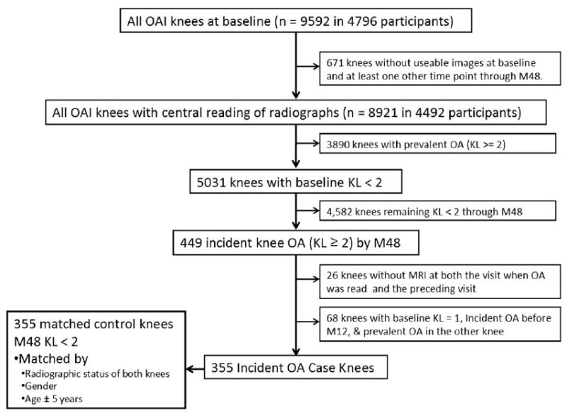
Methods: We studied 355 knees from the Osteoarthritis Initiative that developed ROA (Kellgren-Lawrence grade ≥ 2), which were matched with control knees. The MR images were assessed using the semi-quantitative MOAKS system. Conditional logistic regression was applied to estimate risk of incident ROA. Logistic regression was used to assess the risk of worsening cartilage damage in knees with partial meniscectomy that developed ROA.
Results: In the group with incident ROA, 4.4 % underwent partial meniscectomy during the year prior to the case-defining visit, compared with none of the knees that did not develop ROA. All (n = 31) knees that had partial meniscectomy and 58.9 % (n = 165) of the knees with prevalent meniscal damage developed ROA (OR = 2.51, 95 % CI [1.73, 3.64]). In knees that developed ROA, partial meniscectomy was associated with an increased risk of worsening cartilage damage (OR = 4.51, 95 % CI [1.53, 13.33]).
Conclusions: The probability of having had partial meniscectomy was higher in knees that developed ROA. When looking only at knees that developed ROA, partial meniscectomy was associated with greater risk of worsening cartilage damage.
Key points: • Partial meniscectomy is a controversial treatment option for degenerative meniscal tears. • Partial meniscectomy is strongly associated with incident osteoarthritis within 1 year. • Partial meniscectomy is associated with increased risk of worsening cartilage damage.
Keywords: Cartilage loss; MRI; Meniscus; Osteoarthritis; Partial meniscectomy.
Figures
References
-
- Lubowitz JH, Poehling GG. Save the meniscus. Arthroscopy. 2011;27:301–302. - PubMed
-
- Burns TC, Giuliani JR, Svoboda SJ, Owens BD. Meniscus repair and transplantation techniques. J Knee Surg. 2011;24:167–174. - PubMed
-
- Englund M, Lohmander LS. Risk factors for symptomatic knee osteoarthritis fifteen to twenty-two years after meniscectomy. Arthritis Rheum. 2004;50:2811–2819. - PubMed
-
- Englund M, Roemer FW, Hayashi D, Crema MD, Guermazi A. Meniscus pathology, osteoarthritis and the treatment controversy. Nat Rev Rheumatol. 2012;8:412–419. - PubMed
-
- Herrlin SV, Wange PO, Lapidus G, Hallander M, Werner S, Weidenhielm L. Is arthroscopic surgery beneficial in treating non-traumatic, degenerative medial meniscal tears? A five year follow-up. Knee Surg Sports Traumatol Arthrosc. 2013;21:358–364. - PubMed
MeSH terms
Grants and funding
LinkOut - more resources
Full Text Sources
Other Literature Sources
Medical
