. 2016 Apr 27;36(17):4708-18.
doi: 10.1523/JNEUROSCI.1092-15.2016.

Clinically Anxious Individuals Show Disrupted Feedback between Inferior Frontal Gyrus and Prefrontal-Limbic Control Circuit

Affiliations

Clinically Anxious Individuals Show Disrupted Feedback between Inferior Frontal Gyrus and Prefrontal-Limbic Control Circuit

Jiook Cha et al. J Neurosci. .

Abstract

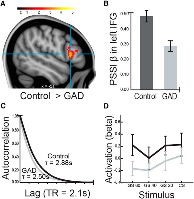
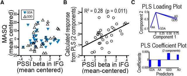
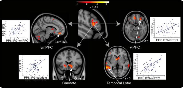
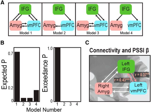
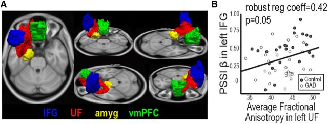
Clinical anxiety is associated with generalization of conditioned fear, in which innocuous stimuli elicit alarm. Using Pavlovian fear conditioning (electric shock), we quantify generalization as the degree to which subjects' neurobiological responses track perceptual similarity gradients to a conditioned stimulus. Previous studies show that the ventromedial prefrontal cortex (vmPFC) inversely and ventral tegmental area directly track the gradient of perceptual similarity to the conditioned stimulus in healthy individuals, whereas clinically anxious individuals fail to discriminate. Here, we extend this work by identifying specific functional roles within the prefrontal-limbic circuit. We analyzed fMRI time-series acquired from 57 human subjects during a fear generalization task using entropic measures of circuit-wide regulation and feedback (power spectrum scale invariance/autocorrelation), in combination with structural (diffusion MRI-probabilistic tractography) and functional (stochastic dynamic causal modeling) measures of prefrontal-limbic connectivity within the circuit. Group comparison and correlations with anxiety severity across 57 subjects revealed dysregulatory dynamic signatures within the inferior frontal gyrus (IFG), which our prior work has linked to impaired feedback within the circuit. Bayesian model selection then identified a fully connected prefrontal-limbic model comprising the IFG, vmPFC, and amygdala. Dysregulatory IFG dynamics were associated with weaker reciprocal excitatory connectivity between the IFG and the vmPFC. The vmPFC exhibited inhibitory influence on the amygdala. Our current results, combined with our previous work across a threat-perception spectrum of 137 subjects and a meta-analysis of 366 fMRI studies, dissociate distinct roles for three prefrontal-limbic regions, wherein the IFG provides evaluation of stimulus meaning, which then informs the vmPFC in inhibiting the amygdala.

Significance statement: Affective neuroscience has generally treated prefrontal regions (orbitofrontal cortex, dorsolateral prefrontal cortex, inferior frontal gyrus, ventromedial prefrontal cortex) equivalently as inhibitory components of the prefrontal-limbic system. Yet research across the anxiety spectrum suggests that the inferior frontal gyrus may have a more complex role in emotion regulation, as this region shows abnormal function in disorders of both hyperarousal and hypoarousal. Using entropic measures of circuit-wide regulation and feedback, in combination with measures of structural and functional connectivity, we dissociate distinct roles for three prefrontal-limbic regions, wherein the inferior frontal gyrus provides evaluation of stimulus meaning, which then informs the ventromedial prefrontal cortex in inhibiting the amygdala. This reconfiguration coheres with studies of conceptual disambiguation also implicating the inferior frontal gyrus.

Keywords: connectivity; control systems; dynamic causal modeling; functional MRI; inferior frontal gyrus; prefrontal cortex.


Figures

Figure 1.
Figure 1.
Fear generalization paradigm. Participants (N = 57; n = 32 individuals with GAD) were instructed that they would be shown a red rectangle, which would be paired with a 50% probability of electric shock to the forearm (CS). During fear conditioning, participants viewed five pseudorandom presentations of the CS, paired with a shock each time, and six alternative stimuli (GS), which varied systematically in width from the CS (±20%, ±40%, ±60%) and indicated no shock (conditioning phase; data not shown). The fMRI task preceded the conditioning phase. During the task, there were 15 pseudo-random presentations of each stimulus, resulting in 120 total trials; the task used a 4–10 s (jittered) interstimulus interval with fixation cross and 2 s stimulus presentation. GS were grouped by similarity to CS (i.e., ±20 were analyzed as one condition, as were ±40 and ±60).
Figure 2.
Figure 2.
During fear generalization, individuals with GAD showed functional dynamics within the IFG that were significantly more chaotic (uncontrolled) than those of healthy controls. We quantified dynamics by power spectrum scale invariance β-signatures, which provide the slope of time-series fit to a power law, as well as autocorrelation lifetime τ, which indicates temporal range of time-series' self-similarity: a measure of memory due to negative feedback within the system (A, B). C, Individuals with GAD exhibited significantly shorter τ (less feedback) than controls ([MNI: −54, 18, 18]; p(uncorrected) = 7 × 10⁻⁵; cluster extent = 57; p(corrected) <0.001) for the same region. D, Activation of the same IFG (from a 6-mm-radius sphere centered on the peak voxel of the PSSI group difference result) showed a significant decrease in individuals with GAD compared with healthy controls. Further investigation revealed that the decreased activation in the IFG was independent from the decreased PSSI β-signatures or autocorrelation lifetime τ.
Figure 3.
Figure 3.
Power law β-signatures of the IFG are associated with symptoms of anxiety and depression. A, Scatter plot between PSSI β values versus GDA and GDD. GDA and GDD were selected as significant predictors of PSSI β (among four subscales of MASQ; see Results) (Watson et al., 1995). B, C, Partial Least Square (PLS) regression showing that two orthogonal factors (components) extracted from MASQ subscales predict PSSI β values. B, Scatter plot between calculated response from PLS model and PSSI β values. C, PLS loading plot and coefficient plot. In the loading plot, two depression symptoms (anhedonic depression, GDD) were significantly separated from two anxiety symptoms (anxious arousal, GDA). In the coefficient plot, anxiety and depression symptoms showed negative and positive coefficients, respectively.
Figure 4.
Figure 4.
IFG PSSI β-signatures correlate with effective connectivity modulated by GS. IFG PSSI β-signatures significantly correlated with the GS-modulated IFG connectivity (differences compared with the baseline) between the IFG-seed region and the vmPFC, ventrolateral prefrontal cortex (vlPFC), caudate, and temporal lobe. Results were corrected for multiple comparison by estimating cluster extents under a voxelwise p threshold of 0.005 that correspond to α of 0.05 (see Materials and Methods); for presentational purposes only, a p threshold of 0.005 with cluster extents of 10 voxels were used here.
Figure 5.
Figure 5.
sDCM suggests the prefrontal-limbic threat circuit consisting of the IFG, vmPFC, and amygdala during fear generalization. A, Model space. B, In Bayesian model selection, Model 1 (a fully connected model) showed the greatest expected probability and exceedance probability. C, Individual variability in bidirectional connectivity between the IFG and vmPFC significantly correlates with IFG PSSI β-signatures, showing stronger connectivity as β-signatures shift to pink noise, and weaker connectivity as β-signatures shift to white noise. **p < 0.01. ***p < 0.001.
Figure 6.
Figure 6.
PSSI β-signatures of the IFG correlate with fiber integrity of the uncinate fasciculus. A, The uncinate fasciculus as a potential anatomical substrate of IFG's interaction with the prefrontal-limbic threat circuit. A representative probabilistic tractography result (posterior distribution) shows the uncinate fasciculus (red) connecting the IFG (blue) with the vmPFC/orbitofrontal cortex (green) and the amygdala (yellow). The subcortical/cortical masks were derived from segmentation and parcellation analyses in Freesurfer (http://freesurfer.net/). B, β-Signatures of the IFG significantly correlate with fiber integrity, indexed by FA, of the uncinate fasiculus. robust reg coeff, Robust regression coefficient.

Similar articles

Cited by

References

    1. Almeida JR, Versace A, Mechelli A, Hassel S, Quevedo K, Kupfer DJ, Phillips ML. Abnormal amygdala-prefrontal effective connectivity to happy faces differentiates bipolar from major depression. Biol Psychiatry. 2009;66:451–459. doi: 10.1016/j.biopsych.2009.03.024. - DOI - PMC - PubMed
    1. American Psychiatric Association. Diagnostic and statistical manual of mental disorder (Ed 4) Washington, DC: American Psychiatric Association Task Force on DSM-IV; 2000.
    1. Aron AR, Fletcher PC, Bullmore ET, Sahakian BJ, Robbins TW. Stop-signal inhibition disrupted by damage to right inferior frontal gyrus in humans. Nat Neurosci. 2003;6:115–116. doi: 10.1038/nn1003. - DOI - PubMed
    1. Bach DR, Seymour B, Dolan RJ. Neural activity associated with the passive prediction of ambiguity and risk for aversive events. J Neurosci. 2009;29:1648–1656. doi: 10.1523/JNEUROSCI.4578-08.2009. - DOI - PMC - PubMed
    1. Barbas H, Pandya DN. Architecture and intrinsic connections of the prefrontal cortex in the rhesus monkey. J Comp Neurol. 1989;286:353–375. doi: 10.1002/cne.902860306. - DOI - PubMed

Publication types