. 2016 Apr 27;36(17):4758-70.
doi: 10.1523/JNEUROSCI.3890-15.2016.

Mutations in Cockayne Syndrome-Associated Genes (Csa and Csb) Predispose to Cisplatin-Induced Hearing Loss in Mice

Affiliations

Mutations in Cockayne Syndrome-Associated Genes (Csa and Csb) Predispose to Cisplatin-Induced Hearing Loss in Mice

Robert N Rainey et al. J Neurosci. .

Abstract

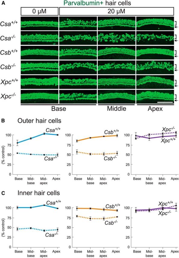
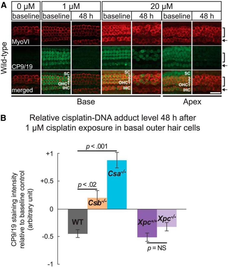
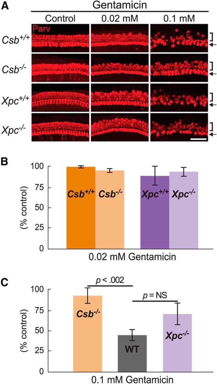
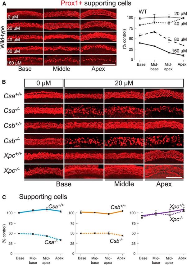
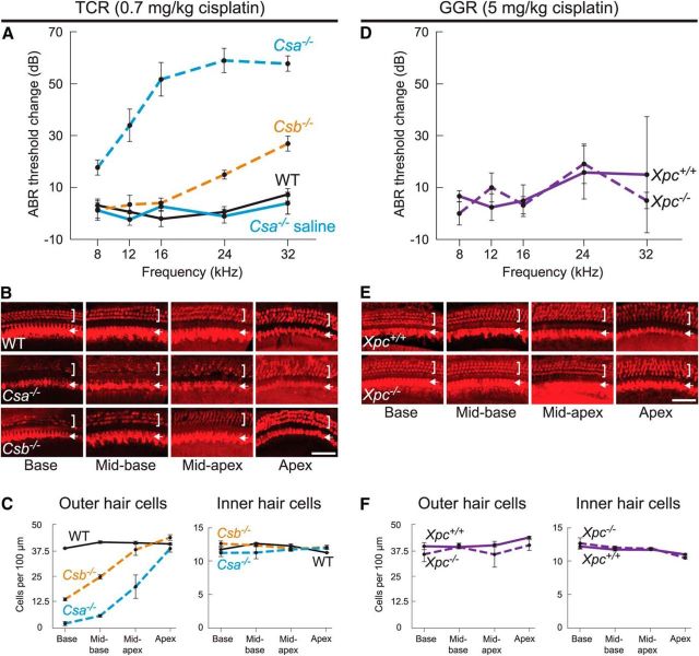
Cisplatin is a common and effective chemotherapeutic agent, yet it often causes permanent hearing loss as a result of sensory hair cell death. The causes of sensitivity to DNA-damaging agents in nondividing cell populations, such as cochlear hair and supporting cells, are poorly understood, as are the specific DNA repair pathways that protect these cells. Nucleotide excision repair (NER) is a conserved and versatile DNA repair pathway for many DNA-distorting lesions, including cisplatin-DNA adducts. Progressive sensorineural hearing loss is observed in a subset of NER-associated DNA repair disorders including Cockayne syndrome and some forms of xeroderma pigmentosum. We investigated whether either of the two overlapping branches that encompass NER, transcription-coupled repair or global genome repair, which are implicated in Cockayne syndrome and xeroderma pigmentosum group C, respectively, modulates cisplatin-induced hearing loss and cell death in the organ of Corti, the auditory sensory epithelium of mammals. We report that cochlear hair cells and supporting cells in transcription-coupled repair-deficient Cockayne syndrome group A (Csa(-/-)) and group B (Csb(-/-)) mice are hypersensitive to cisplatin, in contrast to global genome repair-deficient Xpc(-/-) mice, both in vitro and in vivo We show that sensory hair cells in Csa(-/-) and Csb(-/-) mice fail to remove cisplatin-DNA adducts efficiently in vitro; and unlike Xpc(-/-) mice, Csa(-/-) and Csb(-/-) mice lose hearing and manifest outer hair cell degeneration after systemic cisplatin treatment. Our results demonstrate that Csa and Csb deficiencies predispose to cisplatin-induced hearing loss and hair/supporting cell damage in the mammalian organ of Corti, and emphasize the importance of transcription-coupled DNA repair in the protection against cisplatin ototoxicity.

Significance statement: The utility of cisplatin in chemotherapy remains limited due to serious side effects, including sensorineural hearing loss. We show that mouse models of Cockayne syndrome, a progeroid disorder resulting from a defect in the transcription-coupled DNA repair (TCR) branch of nucleotide excision repair, are hypersensitive to cisplatin-induced hearing loss and sensory hair cell death in the organ of Corti, the mammalian auditory sensory epithelium. Our work indicates that Csa and Csb, two genes involved in TCR, are preferentially required to protect against cisplatin ototoxicity, relative to global genome repair-specific elements of nucleotide excision repair, and suggests that TCR is a major force maintaining DNA integrity in the cochlea. The Cockayne syndrome mice thus represent a model for testing the contribution of DNA repair mechanisms to cisplatin ototoxicity.

Keywords: Cockayne syndrome; DNA repair; cisplatin; hearing loss; ototoxicity; sensory hair cells.


Figures

Figure 1.
Figure 1.
Cochlear sensory outer hair cells, but not the more resistant inner hair cells, are lost in a basal-to-apical gradient in cisplatin-exposed WT organ of Corti in vitro. A–C, Organ of Corti explants from P1 Math1-GFP+ mice were treated with cisplatin at the indicated doses for 2 h and incubated in cisplatin-free medium for 48 h. Whole-mount preparations of 48 h post-treated explants were visualized using Math1-GFP (green), which labels hair cells, and an antibody against parvalbumin (red), a hair cell marker. A, Representative pictures from the basal region show the coincidence of Math1-GFP+ cells with parvalbumin+ hair cells at each cisplatin dose. At 0 μm cisplatin, the explants have three rows of outer hair cells (brackets) and one row of inner hair cells (arrows). B, Representative pictures from basal, middle, and apical regions labeled with Math1-GFP are shown. C, Quantification of the change in outer hair cells and inner hair cells in B along the length of the cochlear explant (base to apex) relative to 48 h untreated control (0 μm cisplatin in B). n = 3 animals for each dose. Error bars indicate SEM. Scale bars: A, B, 100 μm.
Figure 2.
Figure 2.
Transcription-coupled repair, but not global genome repair, is crucial for cochlear hair cell survival in response to cisplatin in vitro. A, Organ of Corti explants from P1 mice defective in TCR (Csa−/−, Csb−/−) or GGR (Xpc−/−) and WT littermates were treated with 20 μm cisplatin for 2 h and incubated in cisplatin-free medium for 48 h. Whole-mount preparations of 48 h post-treated explants were stained with an antibody against parvalbumin (green), a marker of hair cells. Representative pictures from basal, middle, and apical regions are shown. Outer hair cells (brackets) and inner hair cells (arrows) in Csa−/− and Csb−/− mice are hypersensitive to cisplatin throughout the cochlear turn. Scale bar, 100 μm. B, C, Quantification of the change in outer hair cells (B) and inner hair cells (C) along the length of the cochlear explant (base to apex) relative to 48 h untreated control of each genotype (0 μm cisplatin in A). n = 3 animals per experimental group of each genotype. Error bars indicate SEM.
Figure 3.
Figure 3.
Transcription-coupled repair-deficient sensory hair cells are less efficient at removing cisplatin-DNA adducts in vitro. A, Organ of Corti explants from P1 WT mice (C57BL/6J) were treated with 1 or 20 μm cisplatin for 2 h and incubated in cisplatin-free medium for 48 h. Whole-mount preparations were double-labeled with the cisplatin-DNA adduct specific antibody CP9/19 (green) and myosin VI (red), which marks hair cells. Representative pictures of explants fixed at 0 h (20 μm cisplatin) or 6 h (1 μm cisplatin) after the removal of cisplatin are shown as baseline controls (see Materials and Methods). OHC, Outer hair cells (brackets); IHC, inner hair cells (arrows); SC, supporting cells. Scale bar, 25 μm. B, Quantification of staining intensity of cisplatin-DNA adduct antibody in outer sensory hair cells from the basal region of explants defective in TCR (Csa−/−, Csb−/−) or GGR (Xpc−/−) and WT controls treated with 1 μm cisplatin for 2 h at 48 h after treatment relative to baseline (established at 6 h after treatment). WT littermates of Csa and Csb mutant mice were combined and are shown as WT. n ≥ 3 animals in each experimental group. Error bars indicate SEM.
Figure 4.
Figure 4.
Transcription-coupled repair-deficient hair cells are not hypersensitive to gentamicin in vitro. A, Organ of Corti explants from P1 mice defective in TCR (Csb−/−) or GGR (Xpc−/−) and WT littermates were treated with 0.02 mm gentamicin or 0.1 mm gentamicin for 3 h and incubated in gentamicin-free medium for 48 h. Representative pictures from the basal region stained with the hair cell marker parvalbumin are shown. Brackets indicate outer hair cells. Arrows indicate inner hair cells. Scale bar, 50 μm. B, C, Quantification of the change in all hair cells within the basal region of explants treated with 0.02 mm gentamicin (B) or 0.1 mm gentamicin (C) relative to 48 h untreated control of each genotype. WT littermates of Csb and Xpc mutant mice were combined and are shown as WT in C. n = 3 animals per experimental group of each genotype. Error bars indicate SEM.
Figure 5.
Figure 5.
Transcription-coupled repair, but not global genome repair, is crucial for cochlear supporting cell survival in response to cisplatin in vitro. A, Supporting cells are lost in cisplatin-exposed WT organ of Corti in a dose-dependent manner. Organ of Corti explants from P1 Math1-GFP+ mice were treated with cisplatin at the indicated doses as described in Figure 1 and stained with an antibody against Prox1 (red), which marks supporting cells. Quantification of the change in WT supporting cells along the length of the cochlear explant (base to apex) relative to 48 h untreated control (0 μm cisplatin) is shown on the right. n = 3 animals for each dose. B, C, Supporting cells in TCR-deficient Csa−/− and Csb−/− mice, but not in GGR-deficient Xpc−/− mice, are hypersensitive to cisplatin throughout the cochlear turn. B, Organ of Corti explants from P1 mutant mice (Csa−/−, Csb−/−, Xpc−/−) and WT littermates were treated with 20 μm cisplatin as described in Figure 2 and stained with anti-Prox1. C, Supporting cells in B were quantified as in A relative to 48 h untreated control of each genotype (0 μm cisplatin in B). n = 3 animals per experimental group. Scale bars: A, B, 100 μm. A, C, Error bars indicate SEM.
Figure 6.
Figure 6.
Cisplatin-exposed transcription-coupled repair-deficient cochlear hair cells and supporting cells are lost concomitantly through apoptosis in vitro, and the transcription-coupled repair factor CSA is differentially expressed in WT hair cells and supporting cells. A, B, Organ of Corti explants from P1 mice defective in TCR (Csa−/−, Csb−/−) and WT littermates were treated with 20 μm cisplatin as described in Figure 2. Whole-mount preparations and cross sections of 48 h post-treated explants were double-labeled with an antibody against ActCasp3 (red), a marker of apoptosis, and Math1-GFP (green) or p27Kip1 (green) to visualize hair cells (A) or supporting cells (B), respectively. Representative pictures from the basal/mid-basal region of each explant are shown. Brackets indicate outer hair cells. Arrowheads indicate inner hair cells. Supporting cells are indicated as follows: p, Pillar cells; d, Deiters' cells; h, Hensen's cells. C, Parvalbumin+ outer hair cells (OHC) and Prox1+ supporting cells (SC) within the mid-basal region of 20 μm cisplatin-exposed Csb−/− explants were quantified after incubation in cisplatin-free medium for the indicated times relative to 24 or 48 h untreated control. n = 3 animals at each time point. Error bars indicate SEM. D, Adjacent sections through P1 Math1-GFP+ organ of Corti were visualized using Math1-GFP or stained with an antibody against CSA or CSB. High-resolution image shows the overlap of CSA with supporting cells (outlined in dotted white). p, Pillar cells; d1–d3, Deiters' cells. Tectal cells (t1–t2) and undertectal cells (ut) are a subset of Hensen's cells. Arrowheads indicate inner hair cells. Arrows indicate outer hair cells. E, P1 Math1-GFP+ explants were cultured in the presence of 20 ng/ml protein export inhibitor Leptomycin B (+Leptomycin) or its solvent methanol (−Leptomycin) for 16 h and visualized using Math1-GFP or an antibody against CSA. Scale bars: A, B, D, E, 25 μm.
Figure 7.
Figure 7.
Transcription-coupled repair-deficient mice, but not global genome repair-deficient mice, challenged with systemic cisplatin injection present elevated ABR threshold shifts in the high frequencies and outer hair cell loss in a basal-to-apical gradient. A, D, Change in ABR thresholds 2 weeks after 0.7 mg cisplatin/kg body weight treatment in mice defective in TCR (Csa−/−, Csb−/−) and WT littermates (A) or after 5 mg cisplatin/kg body weight treatment in mice defective in GGR (Xpc−/−) and WT littermates (D). WT littermates of Csa and Csb mutant mice were combined. B, E, Whole-mount preparations of the organ of Corti 2 week postcisplatin intraperitoneal injection from A, D were stained with the hair cell marker parvalbumin (red). Representative pictures from each cochlear position are shown. Brackets indicate outer hair cells. Arrows indicate inner hair cells. Scale bars, 50 μm. C, F, Quantification of the change in outer hair cells and inner hair cells in B, E along the length of the cochlear duct (base to apex) is presented as average number of cells per 100 μm in the different cochlear regions. n ≥ 3 animals in each experimental group. Error bars indicate SEM.

Similar articles

Cited by

References

    1. Aamann MD, Hvitby C, Popuri V, Muftuoglu M, Lemminger L, Skeby CK, Keijzers G, Ahn B, Bjorås M, Bohr VA, Stevnsner T. Cockayne Syndrome group B protein stimulates NEIL2 DNA glycosylase activity. Mech Ageing Dev. 2014;135:1–14. doi: 10.1016/j.mad.2013.12.008. - DOI - PMC - PubMed
    1. Andres AL, Gong X, Di K, Bota DA. Low-doses of cisplatin injure hippocampal synapses: a mechanism for ‘chemo’ brain? Exp Neurol. 2014;255:137–144. doi: 10.1016/j.expneurol.2014.02.020. - DOI - PMC - PubMed
    1. Anniko M, Sobin A. Cisplatin: evaluation of its ototoxic potential. Am J Otolaryngol. 1986;7:276–293. doi: 10.1016/S0196-0709(86)80050-3. - DOI - PubMed
    1. Arlett CF, Plowman PN, Rogers PB, Parris CN, Abbaszadeh F, Green MH, McMillan TJ, Bush C, Foray N, Lehmann AR. Clinical and cellular ionizing radiation sensitivity in a patient with xeroderma pigmentosum. Br J Radiol. 2006;79:510–517. doi: 10.1259/bjr/83726649. - DOI - PubMed
    1. Berg RJ, Rebel H, van der Horst GT, van Kranen HJ, Mullenders LH, van Vloten WA, de Gruijl FR. Impact of global genome repair versus transcription-coupled repair on ultraviolet carcinogenesis in hairless mice. Cancer Res. 2000;60:2858–2863. - PubMed

Publication types

Supplementary concepts

LinkOut - more resources