. 2016 Jun 15;129(12):2317-2328.
doi: 10.1242/jcs.179127. Epub 2016 Apr 27.

An initial phase of JNK activation inhibits cell death early in the endoplasmic reticulum stress response

Affiliations

An initial phase of JNK activation inhibits cell death early in the endoplasmic reticulum stress response

Max Brown et al. J Cell Sci. .

Abstract

Accumulation of unfolded proteins in the endoplasmic reticulum (ER) activates the unfolded protein response (UPR). In mammalian cells, UPR signals generated by several ER-membrane-resident proteins, including the bifunctional protein kinase endoribonuclease IRE1α, control cell survival and the decision to execute apoptosis. Processing of XBP1 mRNA by the RNase domain of IRE1α promotes survival of ER stress, whereas activation of the mitogen-activated protein kinase JNK family by IRE1α late in the ER stress response promotes apoptosis. Here, we show that activation of JNK in the ER stress response precedes activation of XBP1. This activation of JNK is dependent on IRE1α and TRAF2 and coincides with JNK-dependent induction of expression of several antiapoptotic genes, including cIap1 (also known as Birc2), cIap2 (also known as Birc3), Xiap and Birc6 ER-stressed Jnk1(-/-) Jnk2(-/-) (Mapk8(-/-) Mapk9(-/-)) mouse embryonic fibroblasts (MEFs) display more pronounced mitochondrial permeability transition and increased caspase 3/7 activity compared to wild-type MEFs. Caspase 3/7 activity is also elevated in ER-stressed cIap1(-/-) cIap2(-/-) and Xiap(-/-) MEFs. These observations suggest that JNK-dependent transcriptional induction of several inhibitors of apoptosis contributes to inhibiting apoptosis early in the ER stress response.

Keywords: Apoptosis; Endoplasmic reticulum; IRE1; JNK; Stress response; Unfolded protein response.


Figures

Fig. 1
Fig. 1
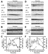
JNK activation precedes activation of XBP1 splicing in MEFs. (A) Kinetics of JNK and eIF2α phosphorylation and (B) XBP1 splicing in MEFs exposed to 1 μM thapsigargin. (C) Quantification of JNK (white circles, solid line, n = 4) and eIF2α (white squares, dotted line) phosphorylation from panel (A) and of XBP1 splicing (black circles, dashed line, n = 2) from panel (B). (D) Kinetics of JNK and eIF2α phosphorylation and (E) XBP1 splicing in MEFs exposed to 10 μg/ml tunicamycin. (F) Quantification of JNK (white circles, solid line, n = 3) and eIF2α (white squares, dotted line) phosphorylation from panel (D) and of XBP1 splicing (black circles, dashed line) from panel (E). p values for comparison of the JNK phosphorylation after addition of the drugs to the cells to JNK phosphorylation in the untreated cells were obtained from an ordinary one way analysis of variance (ANOVA) with Dunnett’s correction for multiple comparisons (Dunnett, 1955; Dunnett, 1964). * - p < 0.05, ** - p < 0.01, *** - p < 0.001, and **** - p < 0.0001. A repeat of the eIF2α Western blots gave qualitatively similar results.
Fig. 2
Fig. 2
IRE1α and TRAF2 are required for the initial phase of JNK activation in MEFs. (A) Kinetics of JNK and eIF2α phosphorylation and (B) XBP1 splicing in ire1α-/- MEFs exposed to 1 μM thapsigargin. For eIF2α phosphorylation qualitatively similar data were obtained in one repeat of the experiment. (C) Quantification of JNK (white circles, solid line, n = 3) and eIF2α (white squares, dotted line) phosphorylation from panel (A) and of XBP1 splicing (black circles, dashed line) from panel (B). (D) Kinetics of JNK and eIF2α phosphorylation and (E) XBP1 splicing in traf2-/- MEFs exposed to 1 μM thapsigargin. eIF2α phosphorylation was expressed relative to the 480 min time point. (F) Quantification of JNK (white circles, solid line, n = 3) and eIF2α phosphorylation (white squares, dotted line, n = 2) from panel (D) and XBP1 splicing (black circles, dashed line) from panel (E). (G) Comparison of phosphorylation of JNK in WT, ire1α-/-, and traf2-/- MEFs before the onset of elevated JNK phosphorylation after 240 min of ER stress in ire1α-/- and traf2-/- MEFs. The bars represent the relative JNK phosphorylation before, 10, 20, 30, 45, 60, 120, and 240 min after addition of thapsigargin to the cells. p values for comparison of JNK phosphorylation in treated cells to the JNK phosphorylation in untreated cells were calculated with an ordinary one way ANOVA with Dunnett’s correction for multiple comparisons.
Fig. 3
Fig. 3
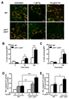
JNK inhibits cell death early in the ER stress response. (A) WT and jnk1-/- jnk2-/- MEFs were treated with 1 μM thapsigargin (Tg) or 10 μg/ml tunicamycin (Tm) for 4 h and stained with JC-1 as described in Materials and Methods. Scale bar – 10 μm. (B, C) Quantification of the confocal fluorescence microscopy data shown in panel A for (B) thapsigargin- and (C) tunicamycin-treated cells. At least 600 cells were counted for each sample. (D) Combined activities of caspases 3 and 7 in WT and jnk1-/- jnk2-/- MEFs treated for 4 h with 1 or 2 μM thapsigargin (Tg) or (E) 10 μg/ml tunicamycin (Tm). The combined caspase activities are expressed relative to the untreated cells. p values were calculated with an ordinary two way ANOVA with Šidák’s correction for multiple comparisons (Šidák, 1967) (n = 3 for panels D and E).
Fig. 4
Fig. 4
JNK is required for transcriptional induction of antiapoptotic genes early in the ER stress response. (A) cIAP1 (BIRC2), (B) cIAP2 (BIRC3), (C) XIAP (BIRC4), and (D) BIRC6 (BRUCE) steady-state mRNA levels were quantified by RT-qPCR in WT and jnk1-/- jnk2-/- MEFs exposed to 1 μM thapsigargin for the indicated times. The p values for the genotype comparisons of an ordinary two way ANOVA with Šidák’s correction for multiple comparisons are shown (n = 3).
Fig. 5
Fig. 5
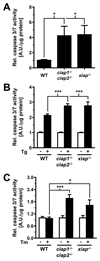
cIAP1, cIAP2, and XIAP protect against apoptosis early in the ER stress response. (A) Combined activities of caspases 3 and 7 in untreated WT, ciap1-/- ciap2-/-, and xiap-/- MEFs and after exposure to (B) 2 μM thapsigargin (Tg) or (C) 10 μg/ml tunicamycin (Tm) for 4 h. p values were calculated with an ordinary two way ANOVA with Dunnett’s (panel A) or Tukey’s (Tukey, 1949) (panels B and C) correction for multiple comparisons (n = 3).
Fig. 6
Fig. 6
Immediately activated JNK localizes to the cytosol during ER stress. Serum-starved Hep G2 cells were treated for 45 min with 1 μM thapsigargin or left untreated before isolation of the cytosolic and nuclear fractions. The cytosolic (C) and nuclear (N) fractions were analysed by Western blotting. The asterisk (*) indicates a non-specific band recognised by the anti-emerin antibody. Emerin was used as a nuclear marker and GAPDH as a cytoplasmic marker. The experiment was repeated once with qualitatively similar results.

References

    1. Amato PA, Unanue ER, Taylor DL. Distribution of actin in spreading macrophages: a comparative study on living and fixed cells. J Cell Biol. 1983;96:750–61. - PMC - PubMed
    1. Ankarcrona M, Dypbukt JM, Bonfoco E, Zhivotovsky B, Orrenius S, Lipton SA, Nicotera P. Glutamate-induced neuronal death: a succession of necrosis or apoptosis depending on mitochondrial function. Neuron. 1995;15:961–73. - PubMed
    1. Arshad M, Ye Z, Gu X, Wong CK, Liu Y, Li D, Zhou L, Zhang Y, Bay WP, Yu VC, et al. RNF13, a RING finger protein, mediates endoplasmic reticulum stress-induced apoptosis through the IRE1alpha/JNK pathway. J Biol Chem. 2013;288:8726–36. - PMC - PubMed
    1. Bains W, Ponte P, Blau H, Kedes L. Cardiac actin is the major actin gene product in skeletal muscle cell differentiation in vitro. Mol Cell Biol. 1984;4:1449–53. - PMC - PubMed
    1. Blau HM, Pavlath GK, Hardeman EC, Chiu CP, Silberstein L, Webster SG, Miller SC, Webster C. Plasticity of the differentiated state. Science. 1985;230:758–66. - PubMed

MeSH terms

Substances