. 2016 Jul;152(1):194-204.
doi: 10.1093/toxsci/kfw081. Epub 2016 Apr 27.

Copper Exposure Perturbs Brain Inflammatory Responses and Impairs Clearance of Amyloid-Beta

Affiliations

Copper Exposure Perturbs Brain Inflammatory Responses and Impairs Clearance of Amyloid-Beta

Masashi Kitazawa et al. Toxicol Sci. 2016 Jul.

Abstract

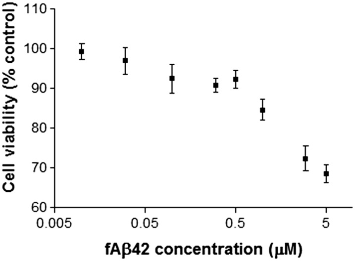
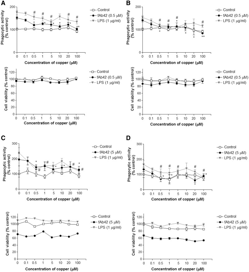
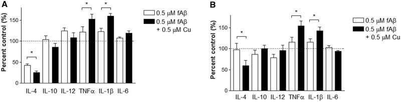
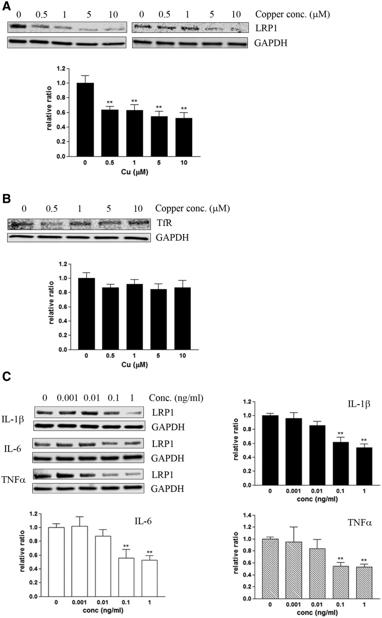
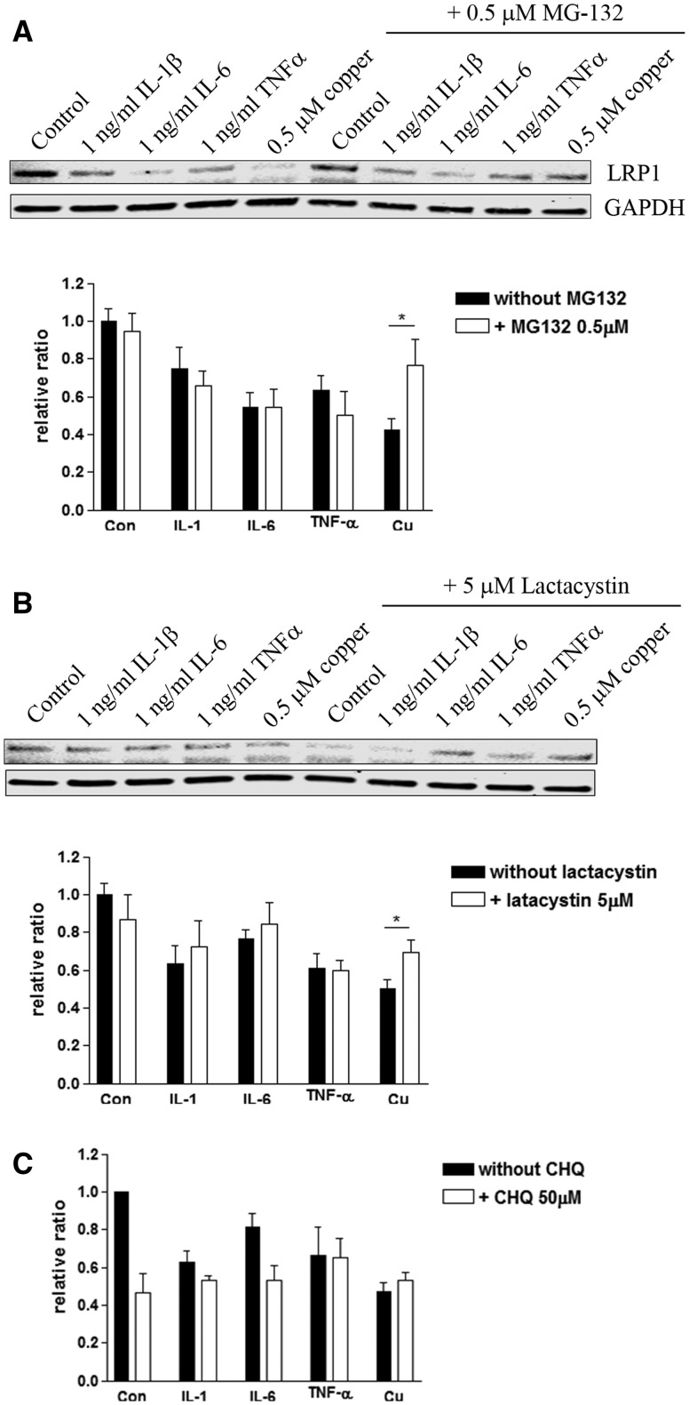
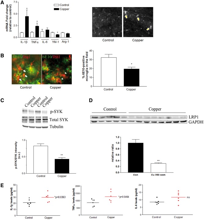
Copper promotes a toxic buildup of amyloid-beta (Aβ) and neurofibrillary tangle pathology in the brain, and its exposure may increase the risk for Alzheimer's disease (AD). However, underlying molecular mechanisms by which copper triggers such pathological changes remain largely unknown. We hypothesized that the copper exposure perturbs brain inflammatory responses, leading to impairment of Aβ clearance from the brain parenchyma. Here, we investigated whether copper attenuated Aβ clearance by microglial phagocytosis or by low-density lipoprotein-related receptor protein-1 (LRP1) dependent transcytosis in both in vitro and in vivo When murine monocyte BV2 cells were exposed to copper, their phagocytic activation induced by fibrillar Aβ or LPS was significantly reduced, while the secretion of pro-inflammatory cytokines, such as IL-1β, TNF-α, and IL-6, were increased. Interestingly, not only copper itself but also IL-1β, IL-6, or TNF-α were capable of markedly reducing the expression of LRP1 in human microvascular endothelial cells (MVECs) in a concentration-dependent manner. While copper-mediated downregulation of LRP1 was proteasome-dependent, the cytokine-induced loss of LRP1 was proteasome- or lysosome-independent. In the mouse model, copper exposure also significantly elevated neuroinflammation and downregulated LRP1 in the brain, consistent with our in vitro results. Taken together, our findings support the pathological impact of copper on inflammatory responses and Aβ clearance in the brain, which could serve as key mechanisms to explain, in part, the copper exposure as an environmental risk factor for AD.

Keywords: Alzheimer’s disease; LRP1; copper; cytokines; inflammation; microglia; phagocytosis.


Figures

FIG. 1
FIG. 1
Fibrillar Aβ induces acute cytotoxicity to BV2 cells. BV2 cells were exposed to a range of concentrations (0–5 µM) of synthetic fibrillar Aβ1-42 (fAβ) for 1 h in a serum-free condition. Acute fAβ toxicity was determined by the MTT assay. Each plot represents mean ± SEM of 3–4 separate experiments in triplicates (n = 9–12).
FIG. 2
FIG. 2
Copper exposure significantly inhibits fAβ-induced phagocytosis by BV2 cells. BV2 cells were exposed to various concentrations (0, 0.1, 0.5, 1, 5, 10, 20, 100 µM) of copper for 3 or 24 h followed by 1-h exposure to 0.5 or 5 µM fAβ or 1 µg/ml LPS to stimulate phagocytosis. A, Phagocytic activation of BV2 cells and toxicity assay following the 3-h copper exposure and 0.5 µM fAβ stimulation. B, Phagocytic activation of BV2 cells and toxicity assay following the 24-h copper exposure and 0.5 µM fAβ stimulation. C, Phagocytic activation of BV2 cells and toxicity assay following the 3-h copper exposure and 5 µM fAβ stimulation. D, Phagocytic activation of BV2 cells and toxicity assay following the 24-h copper exposure and 5 µM fAβ stimulation. Each graph represents mean ± SEM of 3 separate experiments in triplicates (n = 9). Veh = vehicle control (no stimulation). *P < 0.05 or **P < 0.01 compared to the corresponding stimulating agent (fAβ or LPS) alone.
FIG. 3
FIG. 3
Copper exposure suppresses phagocytic markers and inactivates SYK in BV2 cells. A, Phagocytic assay by fluorescent microbeads demonstrates a significant reduction of BV2 cell population that takes up more than 3 beads per cell in the 24-h copper (0.5 µM) exposure followed by 0.5 µM fAβ stimulation for 1 h. Each graph represents mean ± SEM of 3 separate experiments in triplicates (n = 9). *P < 0.05 compared to the fAβ-stimulated group. B, The expression of YM-1 and Arg-1 in 0.5 µM fAβ alone or copper (0.5 µM, 24 h) exposure followed by 0.5 µM fAβ stimulation for 1 h. Each graph represents mean ± S.E.M. of 3 separate experiments in triplicates (n = 9). *P < 0.05 compared to the fAβ-stimulated group. C, Immunoblot analysis of phospho-SYK in BV2 cells. The band intensity of phospho-SYK or total SYK was first normalized by tubulin, then the ratio phospho-SYK/total SYK was calculated. Each graph represents mean ± S.E.M. of 2 separate experiments in triplicates (n = 6). *P < 0.05 or **P < 0.01 compared to the fAβ-stimulated group, #P < 0.05 compared to the control group.
FIG. 4
FIG. 4
Copper exposure activates pro-inflammatory responses in BV2 cells when stimulated with fAβ. Conditioned media were collected and quantitatively analyzed for selected pro- and anti-inflammatory cytokines released from BV2 cells after 1-h fAβ (0.5 µM) stimulation. A, BV2 cells pre-exposed to 0.5 µM copper for 3 h or B, BV2 cells pre-exposed to 0.5 µM copper for 24 h. Each graph represents mean ± S.E.M. of 3 separate experiments in triplicates (n = 9). *P < 0.05 compared to the fAβ-stimulated group without pre-exposure to copper (open bar).
FIG. 5
FIG. 5
Copper and pro-inflammatory cytokines down-regulate LRP1 in microvascular endothelial cells. Human primary microvascular endothelial cells (MVECs) were exposed to copper or human recombinant IL-1β, IL-6 or TNFα for 24 h, and the steady-state levels of LRP1 was quantitatively measured by immunoblot. A, Copper down-regulates LRP1 in MVECs in a concentration-dependent manner. B, Transferrin receptor (TfR) was not altered in MVEC exposed to copper. C, IL-1β, IL-6 or TNFα alone also significantly down-regulates LRP1 in MVECs in a concentration-dependent manner. Each graph represents mean ± S.E.M. of 3 separate experiments (n = 6–9). *P < 0.05 or **P < 0.01 compared to the control group.
FIG. 6
FIG. 6
Cytokine-induced downregulation of LRP1 is not mediated by proteasome degradation. A, MVECs were exposed to copper, IL-1β, IL-6 or TNFα in the presence or absence of proteasome inhibitors, MG-132 (0.5 µM, A) or lactacystin (5 µM, B), for 24 h, and the steady-state levels of LRP1 were measured. The graph represents mean ± S.E.M. of 3 separate experiments (n = 8–11). *P < 0.05 compared to the group without proteasome inhibitor. C, MVECs were exposed to copper, IL-1β, IL-6 or TNFα in the presence or absence of a lysosomal inhibitor, CHQ, for 24 h, and the steady-state levels of LRP1 were measured. The graph represents mean ± S.E.M. of 3 separate experiments in duplicates (n = 6). No significance was observed.
FIG. 7
FIG. 7
Aberrant activation of neuroinflammation in the brain of copper-exposed 3xTg-AD mice. Three months old 3xTg-AD mice were treated with 250 ppm copper-containing drinking water for 3–12 months. A, The mRNA expression of selected cytokines and phagocytosis markers in the brain after the 3 months of control water (n = 10, open bars) or copper exposure (n = 10, closed bars). *P < 0.05 compared to the control group. Representative immunofluorescent staining of Iba1-positive microglia in hippocampus of 3xTg-AD mice received control or copper-containing drinking water for 3 months shows early signs of microglial activation. Arrows indicate the clustering and activating microglia in the absence of Aβ plaques in the copper-exposed mice. B, Representative double immunofluorescent staining of Iba1-positive microglia (red) and 6E10-positive Aβ plaques (green) in hippocampus of 3xTg-AD mice received control or copper-containing drinking water for 9 months. Arrows indicate microglia colocalized with 6E10-positive Aβ, and arrowheads indicate microglia located nearby Aβ plaques but no obvious 6E10-positive Aβ within. The graph represents the percentage of Iba1-positive microglia that colocalize 6E10 positive Aβ. *P < 0.05 compared to the control group (n = 5–8). C, The activation of SYK was determined by immunoblot of phospho-SYK and total SYK in the brain homogenates from 9 months treatment (**P < 0.01 compared to control, n = 4). D, The down-regulation of brain LRP1 was detected in 3xTg-AD mice exposed to copper-containing drinking water for 12 months (**P < 0.01, control n = 5, copper-exposed n = 8). E, Brain cytokine levels were measured by ELISA following 12 months copper exposure (n = 6).

References

    1. Bayer T. A., Schafer S., Simons A., Kemmling A., Kamer T., Tepest R., Eckert A., Schussel K., Eikenberg O., Sturchler-Pierrat C., et al. (2003). Dietary Cu stabilizes brain superoxide dismutase 1 activity and reduces amyloid Abeta production in APP23 transgenic mice. Proc. Natl. Acad. Sci. USA 100, 14187–14192. - PMC - PubMed
    1. Brewer G. J., Kanzer S. H., Zimmerman E. A., Celmins D. F., Heckman S. M., Dick R. (2010). Copper and ceruloplasmin abnormalities in Alzheimer's disease. Am. J. Alzheimer's Dis. Other Dement 25, 490–497. - PMC - PubMed
    1. Burkhead J. L., Gray L. W., Lutsenko S. (2011). Systems biology approach to Wilson's disease. Biometals 24, 455–466. - PMC - PubMed
    1. Chakrabarty P., Jansen-West K., Beccard A., Ceballos-Diaz C., Levites Y., Verbeeck C., Zubair A. C., Dickson D., Golde T. E., Das P. (2010). Massive gliosis induced by interleukin-6 suppresses Abeta deposition in vivo: evidence against inflammation as a driving force for amyloid deposition. Faseb J. 24, 548–559. - PMC - PubMed
    1. Chakrabarty P., Li A., Ceballos-Diaz C., Eddy J. A., Funk C. C., Moore B., DiNunno N., Rosario A. M., Cruz P. E., Verbeeck C., et al. (2015). IL-10 alters immunoproteostasis in APP mice, increasing plaque burden and worsening cognitive behavior. Neuron 85, 519–533. - PMC - PubMed

Publication types

MeSH terms