

Temporal and spatial expression patterns of biomineralization proteins during early development in the stony coral Pocillopora damicornis
- PMID: 27122561
- PMCID: PMC4855386
- DOI: 10.1098/rspb.2016.0322
Temporal and spatial expression patterns of biomineralization proteins during early development in the stony coral Pocillopora damicornis
Abstract
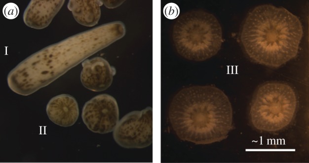
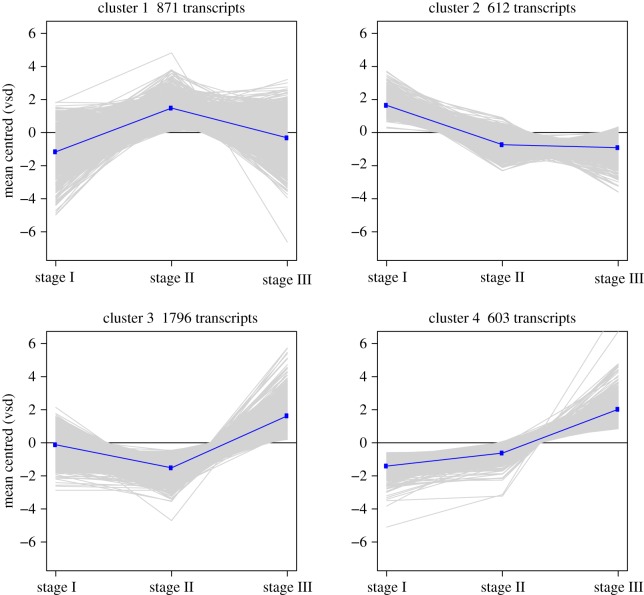
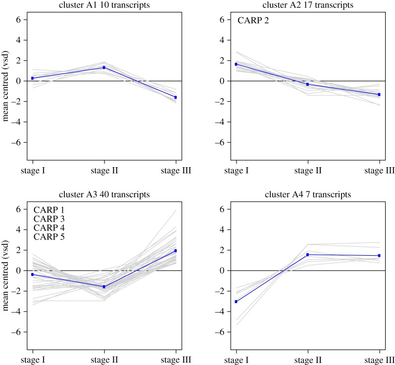
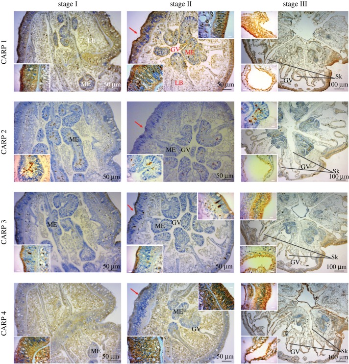
Reef-building corals begin as non-calcifying larvae that, upon settling, rapidly begin to accrete skeleton and a protein-rich skeletal organic matrix that attach them to the reef. Here, we characterized the temporal and spatial expression pattern of a suite of biomineralization genes during three stages of larval development in the reef-building coral Pocillopora damicornis: stage I, newly released; stage II, oral-aborally compressed and stage III, settled and calcifying spat. Transcriptome analysis revealed 3882 differentially expressed genes that clustered into four distinctly different patterns of expression change across the three developmental stages. Immunolocalization analysis further reveals the spatial arrangement of coral acid-rich proteins (CARPs) in the overall architecture of the emerging skeleton. These results provide the first analysis of the timing of the biomineralization 'toolkit' in the early life history of a stony coral.
Keywords: acidic proteins; biomineralization; coral development; gene expression.
© 2016 The Author(s).
Figures

References
-
- Knoll AH. 2003. Biomineralization and evolutionary history. Rev. Miner.Geochem. 54, 329–356. (10.2113/0540329) - DOI
-
- Pochon X, Stat M, Takabayashi M, Chasqui L, Chauka LJ, Logan DDK, Gates RD. 2010. Comparison of endosymbiotic and free-living Symbiodinium (Dinophyceae) diversity in a Hawaiian reef environment. J. Phycol. 46, 53–65. (10.1111/j.1529-8817.2009.00797.x) - DOI
-
- Von Heider A. 1881. Die Gattung Cladocora Ehrenb. Sber. Akad. Wiss. Wien 84, 634–637.
-
- Johnston IS. 1980. The ultrastructure of skeletogensis in hermatypic corals. Int. Rev. Cytol. 67, 171–214. (10.1016/S0074-7696(08)62429-8) - DOI
-
- Tambutte E, Allemand D, Zoccola D, Meibom A, Lotto S, Caminiti N, Tambutte S. 2007. Observations of the tissue–skeleton interface in the scleractinian coral Stylophora pistillata. Coral Reefs 26, 517–529. (10.1007/s00338-007-0263-5) - DOI
Publication types
MeSH terms
Substances
LinkOut - more resources
Full Text Sources
Other Literature Sources
