Case Reports
. 2016 Apr 21:22:342-51.
eCollection 2016.

A novel start codon mutation of the MERTK gene in a patient with retinitis pigmentosa

Affiliations
Case Reports

A novel start codon mutation of the MERTK gene in a patient with retinitis pigmentosa

Worapoj Jinda et al. Mol Vis. .

Abstract

Purpose: Retinitis pigmentosa (RP) is a clinically and genetically heterogeneous group of inherited retinal degenerations characterized by progressive loss of photoreceptor cells and RPE functions. More than 70 causative genes are known to be responsible for RP. This study aimed to identify the causative gene in a patient from a consanguineous family with childhood-onset severe retinal dystrophy.

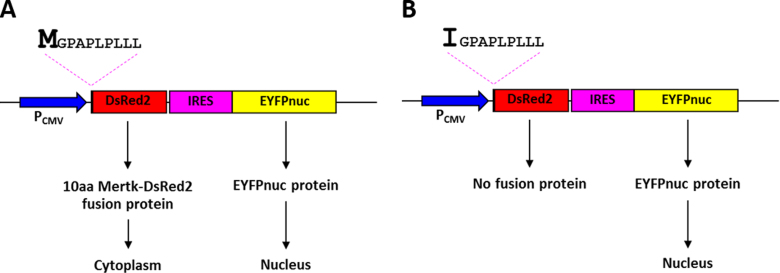
Methods: To identify the defective gene, whole exome sequencing was performed. Candidate causative variants were selected and validated using Sanger sequencing. Segregation analysis of the causative gene was performed in additional family members. To verify that the mutation has an effect on protein synthesis, an expression vector containing the first ten amino acids of the mutant protein fused with the DsRed2 fluorescent protein was constructed and transfected into HEK293T cells. Expression of the fusion protein in the transfected cells was measured using fluorescence microscopy.

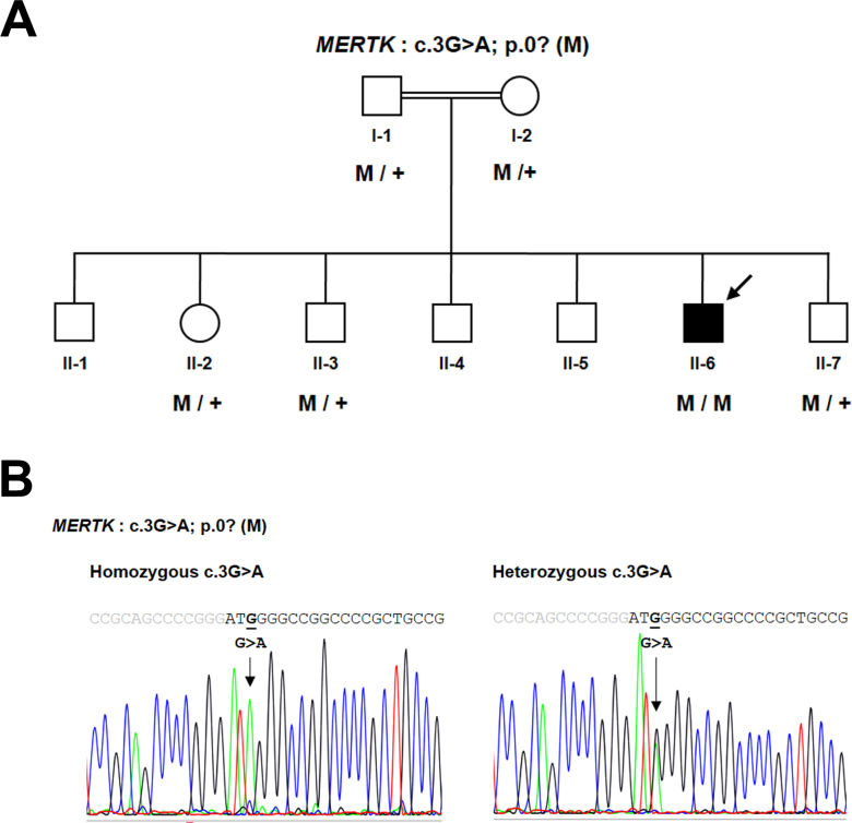
Results: By filtering against public variant databases, a novel homozygous missense mutation (c.3G>A) localized in the start codon of the MERTK gene was detected as a potentially pathogenic mutation for autosomal recessive RP. The c.3G>A mutation cosegregated with the disease phenotype in the family. No expression of the first ten amino acids of the MerTK mutant fused with the DsRed2 fluorescent protein was detected in HEK293T cells, indicating that the mutation affects the translation initiation site of the gene that may lead to loss of function of the MerTK signaling pathway.

Conclusions: We report a novel missense mutation (c.3G>A, p.0?) in the MERTK gene that causes severe vision impairment in a patient. Taken together with previous reports, our results expand the spectrum of MERTK mutations and extend our understanding of the role of the MerTK protein in the pathogenesis of retinitis pigmentosa.


Figures

Figure 1
Figure 1
Fundus photographs, FAF, and OCT results of the patient. Color montage fundus photographs of the right eye (A) and the left eye (B) of the patient at age 35 years. Fundus examination demonstrated changes typical of retinitis pigmentosa (RP) in both eyes, including pale optic discs with attenuated retinal vessels, generalized pigmentary granularity, moderate bone-like spicules in four quadrants, and macular atrophy with pigment accumulation. Fundus autofluorescence (FAF) imaging of the right eye (C) and the left eye (D) showed bilateral decreased autofluorescence corresponding to the areas of pigment accumulation in the center of the macula. These areas were encircled by spotty autofluorescence along the temporal vascular arcades. The optical coherence tomography (OCT) scans of the right eye (E) and the left eye (F) revealed decreased macular thickness.
Figure 2
Figure 2
Pedigree and DNA sequence chromatogram of the family. A: Pedigree of the consanguineous family who participated in this study. Genotype data are presented below the patient and members, where applicable, of his family. The filled symbol with an arrow indicates the index case. Squares: males. Circles: females. Normal alleles are indicated by “+.” The potentially pathogenic mutation, p.0?, is indicated by “M.” B: Sequence analysis of the MERTK gene revealed a missense variant, c.3G>A, located in the start codon of the MerTK protein, homozygous in the patient and heterozygous in other family members. The gray and black letters indicate the 5′ untranslated region (UTR) and exon 1 of the MERTK gene, respectively.
Figure 3
Figure 3
Schematic diagram depicting the cloning strategy for construction of the vectors employed in this study. A: pcDNA3-IRES-EYFPnuc. B: pcDNA3–10 aa MerTK Wt_DsRed2-IRES-EYFPnuc. C: pcDNA3–10 aa MerTK Mut_DsRed2-IRES-EYFPnuc. Blue boxes indicate the cytomegalovirus (CMV) promoter (PCMV) sequence, pink boxes indicate the internal ribosome entry site (IRES) sequence, yellow boxes indicate the enhanced yellow fluorescent protein with nuclear localization signal (EYFPnuc) sequence, red boxes indicate the DsRed2 sequence, and the black arrow indicates the 30 bp MERTK sequence that is a different nucleotide at position +3: (B) G in the Wt vector causing codon 1 to encode Met, and (C) A in the Mut vector causing codon 1 to encode Ile.
Figure 4
Figure 4
Schematic representations of the pcDNA3–10 aa MerTK Wt and Mut-DsRed2-IRES-EYFPnuc vectors and their expected protein expression patterns. The drawings illustrate the pcDNA3–10 aa MerTK Wt-DsRed2-IRES-EYFPnuc vector (A) and the pcDNA3–10 aa MerTK Mut-DsRed2-IRES-EYFPnuc vector (B). Blue arrows indicate the cytomegalovirus (CMV) promoter, which efficiently drives the expression of the bicistronic RNA. The pink dashed lines indicate the first ten amino acid (aa) sequence of the MerTK protein that is a different amino acid at codon 1: Met (M) in 10 aa MerTK Wt (A) and Ile (I) in 10 aa MerTK Mut (B). The red boxes correspond to the DsRed2 sequence, and the pink and yellow boxes indicate the IRES-EYFPnuc sequence. Proteins produced from the wild-type construct vector are predicted to be expressed in the nucleus and the cytoplasm, while protein expression from the mutant construct vector is predicted to be found only in the nucleus.
Figure 5
Figure 5
Identification of fluorescence proteins in HEK293T cells. Protein expression of the Wt or Mut vectors was examined 48 h after transfection. Images were captured using a 40X objective lens. AC: Images of HEK293T cells transfected with the Wt construct encoding an enhanced green fluorescent protein that localizes to the nucleus (A), the fusion protein of 10 aa MerTK Wt-DsRed2 is present in the cytoplasm (B). Merged images (A and B) confirm the fusion protein of the Wt construct is expressed only in the cytoplasm (C). D–F: Images of HEK293T cells transfected with the Mut construct encoding an enhanced green fluorescent protein that localizes to the nucleus (D). E: No fluorescence fusion protein from the Mut construct was detectable in the cytoplasm. The merged images (D and E) indicate no fluorescence fusion protein expressed, except the enhanced green fluorescent protein that was observed in the nucleus (F).
Figure 6
Figure 6
Schematic representation of the MERTK gene at the transcript level and functional domains of the MerTK protein. The MERTK gene consists of 19 exons. Mutations in the MERTK gene are distributed along the entire gene. MerTK is a 999 amino acid protein that contains several conserved domains: Ig-like C2-type 1 (IgL1), Ig-like C2-type 2 (IgL2), fibronectin type-III 1 (FB1), fibronectin type-III 2 (FB2), transmembrane (TM), and protein kinase (PK). Numbers under the protein line indicate the boundaries of each domain. Nucleotide 1 is A of the ATG initiation codon according to RefSeq NM_006343.2. The amino acid residues are numbered according to RefSeq NP_006334.2, starting at the initiator methionine residue.

References

    1. Anasagasti A, Irigoyen C, Barandika O, Lopez de Munain A, Ruiz-Ederra J. Current mutation discovery approaches in Retinitis Pigmentosa. Vision Res. 2012;75:117–29. http://www.ncbi.nlm.nih.gov/entrez/query.fcgi?cmd=Retrieve&db=PubMed&lis... - PubMed
    1. Tsujikawa M, Wada Y, Sukegawa M, Sawa M, Gomi F, Nishida K, Tano Y. Age at onset curves of retinitis pigmentosa. Arch Ophthalmol. 2008;126:337–40. http://www.ncbi.nlm.nih.gov/entrez/query.fcgi?cmd=Retrieve&db=PubMed&lis... - PubMed
    1. Strauss O. The retinal pigment epithelium in visual function. Physiol Rev. 2005;85:845–81. http://www.ncbi.nlm.nih.gov/entrez/query.fcgi?cmd=Retrieve&db=PubMed&lis... - PubMed
    1. Linger RM, Keating AK, Earp HS, Graham DK. TAM receptor tyrosine kinases: biologic functions, signaling, and potential therapeutic targeting in human cancer. Adv Cancer Res. 2008;100:35–83. http://www.ncbi.nlm.nih.gov/entrez/query.fcgi?cmd=Retrieve&db=PubMed&lis... - PMC - PubMed
    1. D’Cruz PM, Yasumura D, Weir J, Matthes MT, Abderrahim H, LaVail MM, Vollrath D. Mutation of the receptor tyrosine kinase gene Mertk in the retinal dystrophic RCS rat. Hum Mol Genet. 2000;9:645–51. http://www.ncbi.nlm.nih.gov/entrez/query.fcgi?cmd=Retrieve&db=PubMed&lis... - PubMed

Publication types

MeSH terms

LinkOut - more resources