doi: 10.14245/kjs.2016.13.1.37.
Epub 2016 Mar 31.
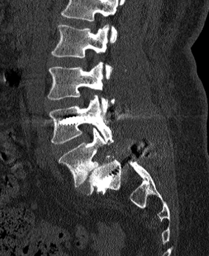
Enlargement of Neural Foramina and Dynamic Stabilization in Spondylolisthesis without Restoring the Alignment: Technical Note
Affiliations
- PMID: 27123030
- PMCID: PMC4844660
- DOI: 10.14245/kjs.2016.13.1.37
Item in Clipboard
Enlargement of Neural Foramina and Dynamic Stabilization in Spondylolisthesis without Restoring the Alignment: Technical Note
Korean J Spine.
2016 Mar.
Abstract
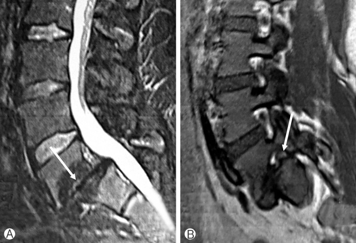
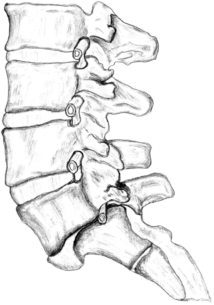
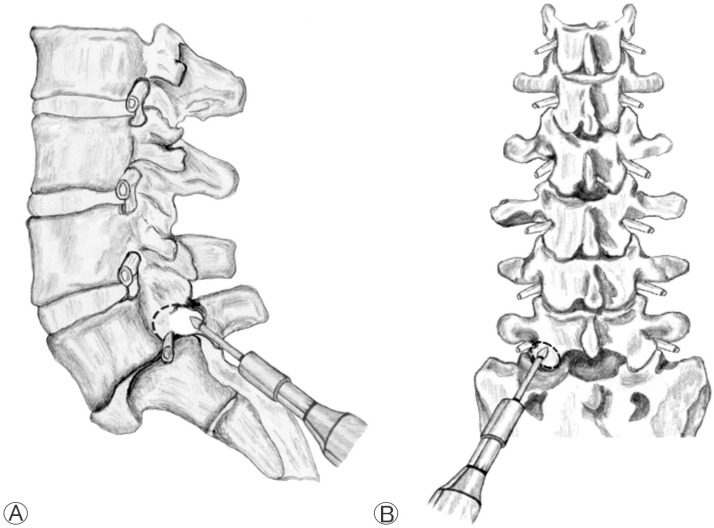
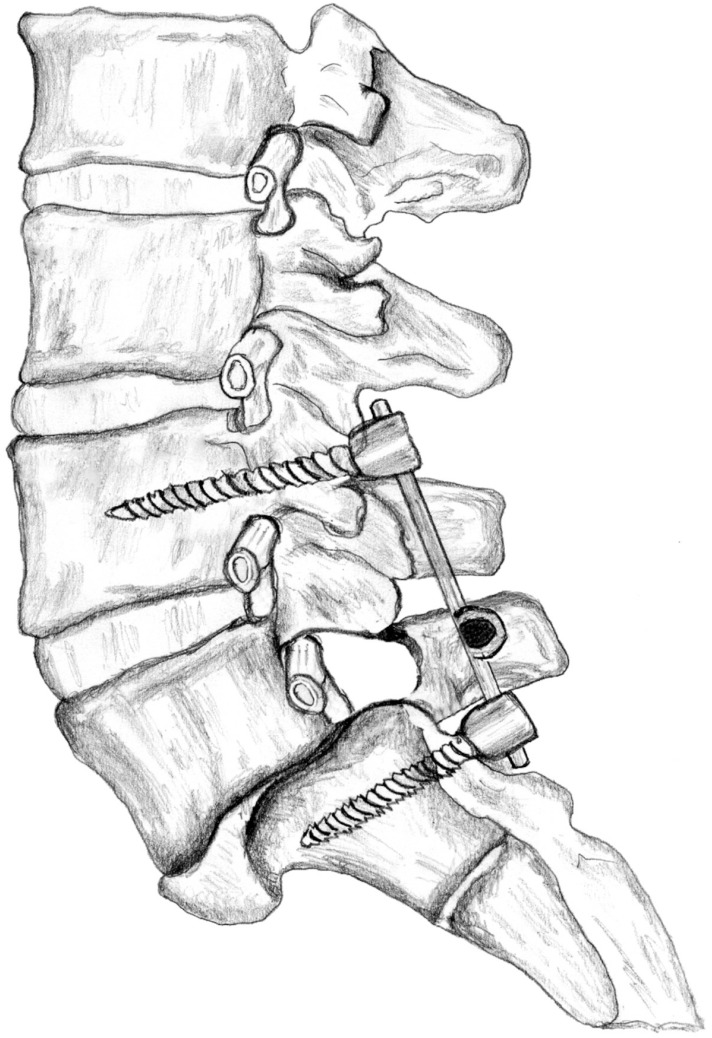
It is well known that the cause of radiculopathy is the compression of the nerve root within the foramina which is narrowed secondary to sliding of the corpus and reduced disc height. In some patients, unroofing the foramen does not resolve this problem. We described a new decompression technique using pedicle removal and transpedicular dynamic instrumentation to stabilization the spine. We performed this operation in 2 patients and achieved very good results.
Keywords: Dynamic stabilization; Lumbar pedicle; Neural foraminal stenosis; Spondylolisthesis.
Conflict of interest statement
Figures
References
-
- Arts M, Pondaag W, Peul W, Thomeer R. Nerve root decompression without fusion in spondylolytic spondylolisthesis: long-term results of Gill's procedure. Eur Spine J. 2006;15:1455–1463. - PubMed
-
- Arts MP, Verstegen MJ, Brand R, Koes BW, van den Akker ME, Peul WC. Cost-effectiveness of decompression according to Gill versus instrumented spondylodesis in the treatment of sciatica due to low grade spondylolytic spondylolisthesis: a pro spective randomised controlled trial [NTR1300] BMC Musculoskelet Disord. 2008;9:128. - PMC - PubMed
-
- Gıll GG, Mannıng JG, Whıte HL. Surgical treatment of spondylolisthesis without spine fusion; excision of the loose lamina with decompression of the nerve roots. J Bone Joint Surg Am. 1955;37-A:493–520. - PubMed
-
- Gill GG, White HL. Surgical treatment of spondylolisthesis without spine fusion. a long term follow-up of operated cases. Acta Orthop Scand Suppl. 1965;(Suppl 85):5–99. - PubMed
LinkOut - more resources
Full Text Sources
Other Literature Sources