. 2016:2016:9438650.
doi: 10.1155/2016/9438650. Epub 2016 Mar 31.

Electroacupuncture Attenuates Cerebral Ischemia and Reperfusion Injury in Middle Cerebral Artery Occlusion of Rat via Modulation of Apoptosis, Inflammation, Oxidative Stress, and Excitotoxicity

Affiliations

Electroacupuncture Attenuates Cerebral Ischemia and Reperfusion Injury in Middle Cerebral Artery Occlusion of Rat via Modulation of Apoptosis, Inflammation, Oxidative Stress, and Excitotoxicity

Mei-Hong Shen et al. Evid Based Complement Alternat Med. 2016.

Abstract

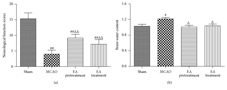
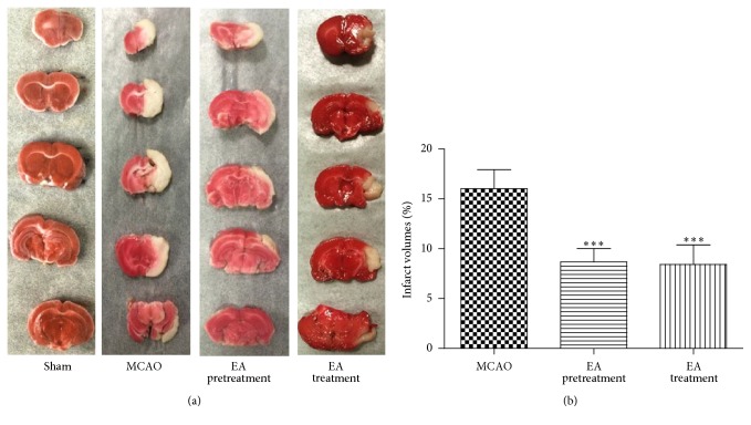
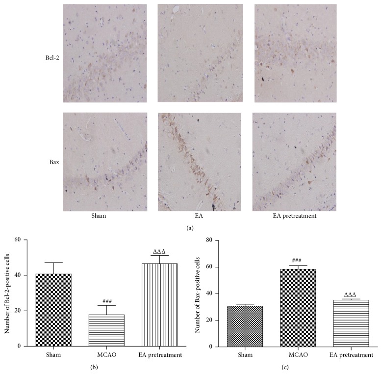
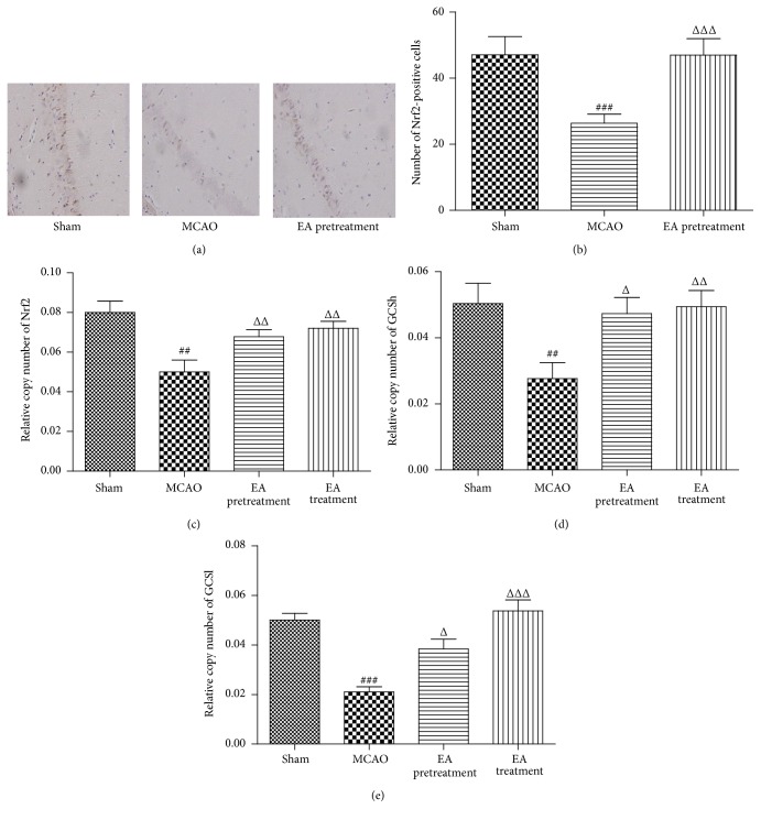
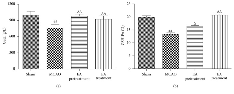
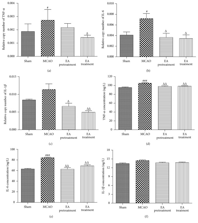
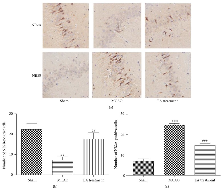
Electroacupuncture (EA) has several properties such as antioxidant, antiapoptosis, and anti-inflammatory properties. The current study was to investigate the effects of EA on the prevention and treatment of cerebral ischemia-reperfusion (I/R) injury and to elucidate possible molecular mechanisms. Sprague-Dawley rats were subjected to middle cerebral artery occlusion (MCAO) for 2 h followed by reperfusion for 24 h. EA stimulation was applied to both Baihui and Dazhui acupoints for 30 min in each rat per day for 5 successive days before MCAO (pretreatment) or when the reperfusion was initiated (treatment). Neurologic deficit scores, infarction volumes, brain water content, and neuronal apoptosis were evaluated. The expressions of related inflammatory cytokines, apoptotic molecules, antioxidant systems, and excitotoxic receptors in the brain were also investigated. Results showed that both EA pretreatment and treatment significantly reduced infarct volumes, decreased brain water content, and alleviated neuronal injury in MCAO rats. Notably, EA exerts neuroprotection against I/R injury through improving neurological function, attenuating the inflammation cytokines, upregulating antioxidant systems, and reducing the excitotoxicity. This study provides a better understanding of the molecular mechanism underlying the traditional use of EA.


Figures

Figure 1
Figure 1
EA improves the neurological function scores and attenuates brain water content in MCAO rats. (a) Neurological function scores at 24 h after reperfusion (n = 6 animals per group). Rats receiving EA pretreatment and EA treatment showed significant improvement in neurological function compared with the MCAO model groups. (b) Both EA pretreatment and EA treatment significantly reduced brain water weight compared with the MCAO group (n = 6 animals per group). Data are represented as mean ± SEM. # P < 0.05 and ## P < 0.01 versus Sham; Δ P < 0.05 and ΔΔ P < 0.01 versus MCAO. EA, electroacupuncture; MCAO, middle cerebral artery occlusion.
Figure 2
Figure 2
Observation of infarction volume: (a) the representative 2,3,5-triphenyltetrazolium chloride (TTC) staining (n = 6 animals per group), ischemic area being white and intact area stained red. (b) Percentage of infarct volume of the cerebral infarct in the rat brain (n = 6 animals per group). Brain tissues displayed obvious infarction in MCAO group compared to Sham-operated group. Both EA pretreatment and EA treatment groups showed a tendency of decrease in infarction volume compared to MCAO group. Results are expressed as mean ± SEM. ∗∗∗ P < 0.001 versus MCAO.
Figure 3
Figure 3
Histological examination of the ischemic cortex tissues of rats was evaluated by HE staining and TEM analysis. (a) Representative images of HE staining performed on sections from the ischemic cortex at 24 h after reperfusion in Sham, MCAO, EA pretreatment, and EA treatment groups (n = 6 animals per group). Ischemic cortex sections obtained from injured cerebral hemispheres were stained with haematoxylin and eosin and observed using Olympus microscope (×400). (b) Ultrastructure changes by transmission electron microscope (×5800). Graphs showing the ultrastructural changes of pyramidal cells, astrocyte, and blood brain barrier (BBB) in different groups (n = 6 animals per group).
Figure 4
Figure 4
Effects of EA on the expression of Bax and Bcl-2 were investigated by immunohistochemistry assay after MCAO. (a–c) Immunohistochemistry staining for Bax or Bcl-2-positive cells in hippocampal CA1 region in different groups (n = 6 animals per group) (×400). Data represent mean ± SEM. ### P < 0.001 versus Sham and ΔΔΔ P < 0.001 versus MCAO.
Figure 5
Figure 5
Effects of EA on the expression of Nrf2 after MCAO. (a and b) Immunohistochemistry staining for Nrf2-positive cells in hippocampal CA1 region in different groups (n = 6 animals per group) (×400). (c–e) Analysis of Nrf2, GCSh, and GCSl mRNA expression levels in cortex by real-time PCR. GAPDH was used as an internal control (n = 8 animals per group). Data represent mean ± SEM. ## P < 0.01, and ### P < 0.001 versus Sham and Δ P < 0.05, ΔΔ P < 0.01, and ΔΔΔ P < 0.001 versus MCAO.
Figure 6
Figure 6
Effects of EA on the activities of GSH and GSH-Px. Treatment with EA significantly increased GSH (a) and GSH-Px (b) activities in serum compared with the MCAO group (n = 8 animals per group). Data represent mean ± SEM. ## P < 0.01 versus Sham and Δ P < 0.05 and ΔΔ P < 0.01 versus MCAO.
Figure 7
Figure 7
Effects of EA on the levels of TNF-α, IL-6, and IL-1β in ischemic cortex and serum after MCAO. (a–c) The levels of TNF-α, IL-6, and IL-1β mRNA in cortex were determined by real-time PCR (n = 8 animals per group). GAPDH was used as an internal control. (d–f) Protein concentrations of TNF-α, IL-6, and IL-1β in the serum in different groups (n = 8 animals per group). Data represent mean ± SEM. ### P < 0.001 versus Sham and Δ P < 0.05 and ΔΔ P < 0.01 versus MCAO. # P < 0.05 versus Sham.
Figure 8
Figure 8
Effects of EA on expression of NR2A and NR2B after MCAO. (a–c) Immunohistochemistry staining for NR2A- and NR2B-positive cells in hippocampal CA1 region in different groups (×400) (n = 6 animals per group). Data represent mean ± SEM. ∗∗ P < 0.01 and ∗∗∗ P < 0.001 versus Sham and ## P < 0.01 and ### P < 0.001 versus MCAO.

Similar articles

Cited by

References

    1. Tsai C.-F., Thomas B., Sudlow C. L. M. Epidemiology of stroke and its subtypes in Chinese vs white populations. Neurology. 2013;81(3):264–272. doi: 10.1212/WNL.0b013e31829bfde3. - DOI - PMC - PubMed
    1. Goldstein L. B., Adams R., Becker K., et al. Primary prevention of ischemic stroke: a statement for healthcare professionals from the stroke council of the American Heart Association. Stroke. 2001;32(1):280–299. doi: 10.1161/01.str.32.1.280. - DOI - PubMed
    1. Allen C. L., Bayraktutan U. Oxidative stress and its role in the pathogenesis of ischaemic stroke. International Journal of Stroke. 2009;4(6):461–470. doi: 10.1111/j.1747-4949.2009.00387.x. - DOI - PubMed
    1. Broughton B. R. S., Reutens D. C., Sobey C. G. Apoptotic mechanisms after cerebral ischemia. Stroke. 2009;40(5):e331–e339. doi: 10.1161/strokeaha.108.531632. - DOI - PubMed
    1. Iadecola C., Anrather J. The immunology of stroke: from mechanisms to translation. Nature Medicine. 2011;17(7):796–808. doi: 10.1038/nm.2399. - DOI - PMC - PubMed

LinkOut - more resources